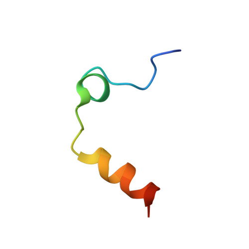
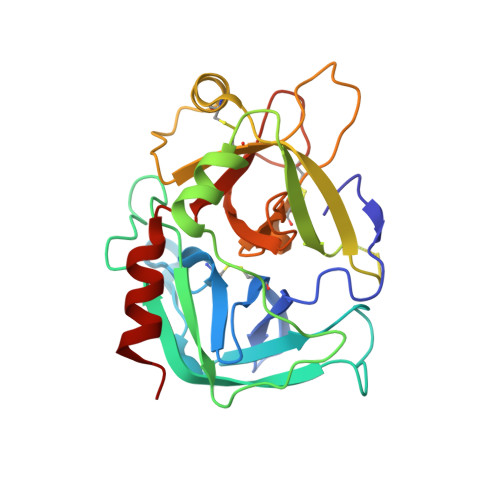
3QGN

The allosteric E*-E equilibrium is a key property of the trypsin fold


Experimental Data Snapshot

  • Method: X-RAY DIFFRACTION
  • Resolution: 2.10 Å
  • R-Value Free: 0.239 
  • R-Value Work: 0.189 
  • R-Value Observed: 0.191 

Starting Model: experimental
View more details

wwPDB Validation   3D Report Full Report


Ligand Structure Quality Assessment 


This is version 1.6 of the entry. See complete history


Literature

Crystallographic and kinetic evidence of allostery in a trypsin-like protease.

Niu, W.Chen, Z.Gandhi, P.S.Vogt, A.D.Pozzi, N.Pelc, L.A.Zapata, F.Di Cera, E.

(2011) Biochemistry 50: 6301-6307

  • DOI: https://doi.org/10.1021/bi200878c
  • Primary Citation of Related Structures:  
    3QGN, 3S7H, 3S7K

  • PubMed Abstract: 

    Protein allostery is based on the existence of multiple conformations in equilibrium linked to distinct functional properties. Although evidence of allosteric transitions is relatively easy to identify by functional studies, structural detection of a pre-existing equilibrium between alternative conformations remains challenging even for textbook examples of allosteric proteins. Kinetic studies show that the trypsin-like protease thrombin exists in equilibrium between two conformations where the active site is either collapsed (E*) or accessible to substrate (E). However, structural demonstration that the two conformations exist in the same enzyme construct free of ligands has remained elusive. Here we report the crystal structure of the thrombin mutant N143P in the E form, which complements the recently reported structure in the E* form, and both the E and E* forms of the thrombin mutant Y225P. The side chain of W215 moves 10.9 Å between the two forms, causing a displacement of 6.6 Å of the entire 215-217 segment into the active site that in turn opens or closes access to the primary specificity pocket. Rapid kinetic measurements of p-aminobenzamidine binding to the active site confirm the existence of the E*-E equilibrium in solution for wild-type and the mutants N143P and Y225P. These findings provide unequivocal proof of the allosteric nature of thrombin and lend strong support to the recent proposal that the E*-E equilibrium is a key property of the trypsin fold.


  • Organizational Affiliation

    Department of Biochemistry and Molecular Biology, Saint Louis University School of Medicine, St. Louis, Missouri 63104, USA.


Macromolecules
Find similar proteins by:  (by identity cutoff)  |  3D Structure
Entity ID: 1
MoleculeChains Sequence LengthOrganismDetailsImage
Thrombin light chain31Homo sapiensMutation(s): 0 
Gene Names: F2
EC: 3.4.21.5
UniProt & NIH Common Fund Data Resources
Find proteins for P00734 (Homo sapiens)
Explore P00734 
Go to UniProtKB:  P00734
PHAROS:  P00734
GTEx:  ENSG00000180210 
Entity Groups  
Sequence Clusters30% Identity50% Identity70% Identity90% Identity95% Identity100% Identity
UniProt GroupP00734
Sequence Annotations
Expand
  • Reference Sequence
Find similar proteins by:  (by identity cutoff)  |  3D Structure
Entity ID: 2
MoleculeChains Sequence LengthOrganismDetailsImage
Thrombin heavy chain259Homo sapiensMutation(s): 1 
Gene Names: F2
EC: 3.4.21.5
UniProt & NIH Common Fund Data Resources
Find proteins for P00734 (Homo sapiens)
Explore P00734 
Go to UniProtKB:  P00734
PHAROS:  P00734
GTEx:  ENSG00000180210 
Entity Groups  
Sequence Clusters30% Identity50% Identity70% Identity90% Identity95% Identity100% Identity
UniProt GroupP00734
Glycosylation
Glycosylation Sites: 1Go to GlyGen: P00734-1
Sequence Annotations
Expand
  • Reference Sequence
Small Molecules
Ligands 3 Unique
IDChains Name / Formula / InChI Key2D Diagram3D Interactions
NAG
Query on NAG

Download Ideal Coordinates CCD File 
D [auth B]2-acetamido-2-deoxy-beta-D-glucopyranose
C8 H15 N O6
OVRNDRQMDRJTHS-FMDGEEDCSA-N
IOD
Query on IOD

Download Ideal Coordinates CCD File 
C [auth A]
E [auth B]
F [auth B]
G [auth B]
H [auth B]
C [auth A],
E [auth B],
F [auth B],
G [auth B],
H [auth B],
I [auth B]
IODIDE ION
I
XMBWDFGMSWQBCA-UHFFFAOYSA-M
CL
Query on CL

Download Ideal Coordinates CCD File 
J [auth B]CHLORIDE ION
Cl
VEXZGXHMUGYJMC-UHFFFAOYSA-M
Experimental Data & Validation

Experimental Data

  • Method: X-RAY DIFFRACTION
  • Resolution: 2.10 Å
  • R-Value Free: 0.239 
  • R-Value Work: 0.189 
  • R-Value Observed: 0.191 
  • Space Group: C 1 2 1
Unit Cell:
Length ( Å )Angle ( ˚ )
a = 122.179α = 90
b = 47.98β = 94.31
c = 52.011γ = 90
Software Package:
Software NamePurpose
CrystalCleardata collection
MOLREPphasing
REFMACrefinement
HKL-2000data reduction
HKL-2000data scaling

Structure Validation

View Full Validation Report


Ligand Structure Quality Assessment 


Entry History 

Deposition Data

Revision History  (Full details and data files)

  • Version 1.0: 2011-07-06
    Type: Initial release
  • Version 1.1: 2011-07-13
    Changes: Version format compliance
  • Version 1.2: 2011-11-16
    Changes: Database references
  • Version 1.3: 2017-11-08
    Changes: Source and taxonomy
  • Version 1.4: 2020-07-29
    Type: Remediation
    Reason: Carbohydrate remediation
    Changes: Data collection, Database references, Derived calculations, Structure summary
  • Version 1.5: 2023-09-13
    Changes: Data collection, Database references, Refinement description, Structure summary
  • Version 1.6: 2024-11-20
    Changes: Structure summary