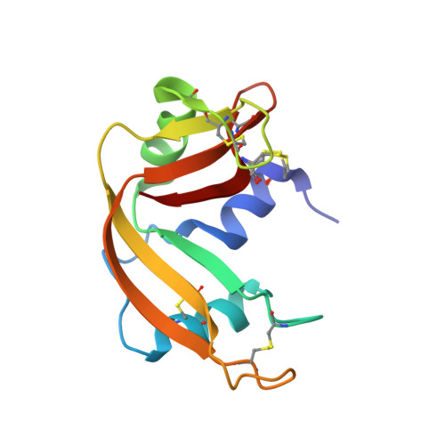
3QL1

Crystal Structure of Ribonuclease A Variant A4C/D83E/V118C


Experimental Data Snapshot

  • Method: X-RAY DIFFRACTION
  • Resolution: 1.29 Å
  • R-Value Free: 0.190 
  • R-Value Work: 0.154 
  • R-Value Observed: 0.155 

Starting Model: experimental
View more details

wwPDB Validation   3D Report Full Report


This is version 1.3 of the entry. See complete history


Literature

Significant stabilization of ribonuclease A by additive effects

Arnold, U.Schopfel, M.

(2012) FEBS J 279: 2508-2519

  • DOI: https://doi.org/10.1111/j.1742-4658.2012.08632.x
  • Primary Citation of Related Structures:  
    3QL1, 3QL2

  • PubMed Abstract: 

    Among the strategies that employ genetic engineering to stabilize proteins, the introduction of disulfide bonds has proven to be a very potential approach. As, however, the replacement of amino acid residues by cysteines and the subsequent formation of the covalent bond can result in a severe deformation of the parental protein structure, the stabilization effect is strongly context dependent. Alternatively, the introduction of charged amino acid residues at the surface, which may result in the formation of extra ionic interactions or hydrogen bonds, provide propitious means for protein stabilization. The generation of an extra disulfide bond between residues 4 and 118 in ribonuclease A had resulted in a stabilization by 6 °C or 7 kJ mol(-1), which was mainly caused by a deceleration of the unfolding reaction [Pecher, P. & Arnold, U. (2009) Biophys Chem, 141, 21-28]. Here, Asp83 was replaced by Glu resulting in a comparable stabilization. Moreover, combination of both mutations led to an additive effect and the resulting ribonuclease A variant (T(m) ~ 76 °C, ΔG° ~ 53 kJ mol(-1)) is the most stable ribonuclease A variant described so far. The analysis of the crystal structure of A4C/D83E/V118C-ribonuclease A reveals the formation of a salt bridge between the γ-carboxyl group of Glu83 and the ε-amino group of Lys104.


  • Organizational Affiliation

    Institute of Biochemistry and Biotechnology, Martin-Luther University Halle-Wittenberg, Halle, Germany. ulrich.arnold@biochemtech.uni-halle.de


Macromolecules
Find similar proteins by:  (by identity cutoff)  |  3D Structure
Entity ID: 1
MoleculeChains Sequence LengthOrganismDetailsImage
Ribonuclease pancreatic124Bos taurusMutation(s): 3 
Gene Names: RNASE1RNS1
EC: 3.1.27.5 (PDB Primary Data), 4.6.1.18 (UniProt)
UniProt
Find proteins for P61823 (Bos taurus)
Explore P61823 
Go to UniProtKB:  P61823
Entity Groups  
Sequence Clusters30% Identity50% Identity70% Identity90% Identity95% Identity100% Identity
UniProt GroupP61823
Sequence Annotations
Expand
  • Reference Sequence
Experimental Data & Validation

Experimental Data

  • Method: X-RAY DIFFRACTION
  • Resolution: 1.29 Å
  • R-Value Free: 0.190 
  • R-Value Work: 0.154 
  • R-Value Observed: 0.155 
  • Space Group: C 1 2 1
Unit Cell:
Length ( Å )Angle ( ˚ )
a = 54.248α = 90
b = 53.781β = 117.95
c = 42.869γ = 90
Software Package:
Software NamePurpose
XSCALEdata scaling
PHASERphasing
REFMACrefinement
PDB_EXTRACTdata extraction
MxCuBEdata collection
XDSdata reduction

Structure Validation

View Full Validation Report


Entry History 

Deposition Data

Revision History  (Full details and data files)

  • Version 1.0: 2012-02-29
    Type: Initial release
  • Version 1.1: 2012-06-13
    Changes: Database references
  • Version 1.2: 2014-05-07
    Changes: Database references
  • Version 1.3: 2023-11-01
    Changes: Data collection, Database references, Derived calculations, Refinement description