3QL3 | pdb_00003ql3

Re-refined coordinates for PDB entry 1RX2


Experimental Data Snapshot

  • Method: X-RAY DIFFRACTION
  • Resolution: 1.80 Å
  • R-Value Free: 
    0.213 (Depositor), 0.220 (DCC) 
  • R-Value Work: 
    0.177 (Depositor), 0.180 (DCC) 
  • R-Value Observed: 
    0.179 (Depositor) 

wwPDB Validation   3D Report Full Report


Ligand Structure Quality Assessment 

Created with Raphaël 2.3.0Worse 01 BetterLigand structure goodness of fit to experimental dataBest fitted NAPClick on this verticalbar to view detailsBest fitted FOLClick on this verticalbar to view details

This is version 1.6 of the entry. See complete history

Re-refinement Note

This entry reflects an alternative modeling of the original data in: 1RX2


Literature

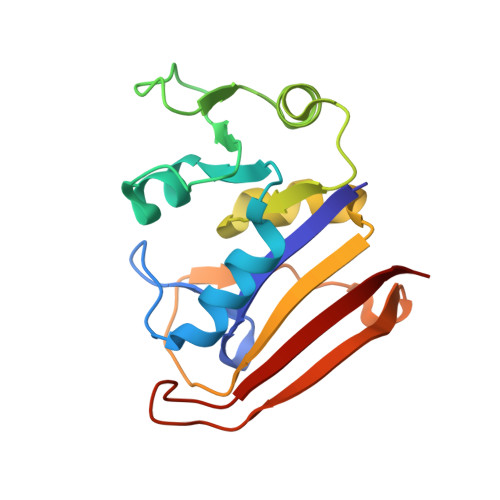
A dynamic knockout reveals that conformational fluctuations influence the chemical step of enzyme catalysis.

Bhabha, G.Lee, J.Ekiert, D.C.Gam, J.Wilson, I.A.Dyson, H.J.Benkovic, S.J.Wright, P.E.

(2011) Science 332: 234-238

  • DOI: https://doi.org/10.1126/science.1198542
  • Primary Citation of Related Structures:  
    3QL0, 3QL3

  • PubMed Abstract: 

    Conformational dynamics play a key role in enzyme catalysis. Although protein motions have clear implications for ligand flux, a role for dynamics in the chemical step of enzyme catalysis has not been clearly established. We generated a mutant of Escherichia coli dihydrofolate reductase that abrogates millisecond-time-scale fluctuations in the enzyme active site without perturbing its structural and electrostatic preorganization. This dynamic knockout severely impairs hydride transfer. Thus, we have found a link between conformational fluctuations on the millisecond time scale and the chemical step of an enzymatic reaction, with broad implications for our understanding of enzyme mechanisms and for design of novel protein catalysts.


  • Organizational Affiliation

    Department of Molecular Biology and Skaggs Institute for Chemical Biology, The Scripps Research Institute, La Jolla, CA 92037, USA.


Macromolecules
Find similar proteins by:  (by identity cutoff)  |  3D Structure
Entity ID: 1
MoleculeChains Sequence LengthOrganismDetailsImage
Dihydrofolate reductase159Escherichia coli K-12Mutation(s): 0 
Gene Names: folAtmrAb0048JW0047
EC: 1.5.1.3
UniProt
Find proteins for P0ABQ4 (Escherichia coli (strain K12))
Explore P0ABQ4 
Go to UniProtKB:  P0ABQ4
Entity Groups  
Sequence Clusters30% Identity50% Identity70% Identity90% Identity95% Identity100% Identity
UniProt GroupP0ABQ4
Sequence Annotations
Expand
  • Reference Sequence
Experimental Data & Validation

Experimental Data

  • Method: X-RAY DIFFRACTION
  • Resolution: 1.80 Å
  • R-Value Free:  0.213 (Depositor), 0.220 (DCC) 
  • R-Value Work:  0.177 (Depositor), 0.180 (DCC) 
  • R-Value Observed: 0.179 (Depositor) 
Space Group: P 21 21 21
Unit Cell:
Length ( Å )Angle ( ˚ )
a = 34.32α = 90
b = 45.51β = 90
c = 98.91γ = 90
Software Package:
Software NamePurpose
REFMACrefinement

Structure Validation

View Full Validation Report


Ligand Structure Quality Assessment 

Created with Raphaël 2.3.0Worse 01 BetterLigand structure goodness of fit to experimental dataBest fitted NAPClick on this verticalbar to view detailsBest fitted FOLClick on this verticalbar to view details

Entry History 

Deposition Data

Revision History  (Full details and data files)

  • Version 1.0: 2011-04-27
    Type: Initial release
  • Version 1.1: 2011-07-13
    Changes: Version format compliance
  • Version 1.2: 2011-08-10
    Changes: Other
  • Version 1.3: 2011-09-28
    Changes: Other
  • Version 1.4: 2012-04-25
    Changes: Other
  • Version 1.5: 2014-11-12
    Changes: Structure summary
  • Version 1.6: 2024-02-21
    Changes: Data collection, Database references, Derived calculations