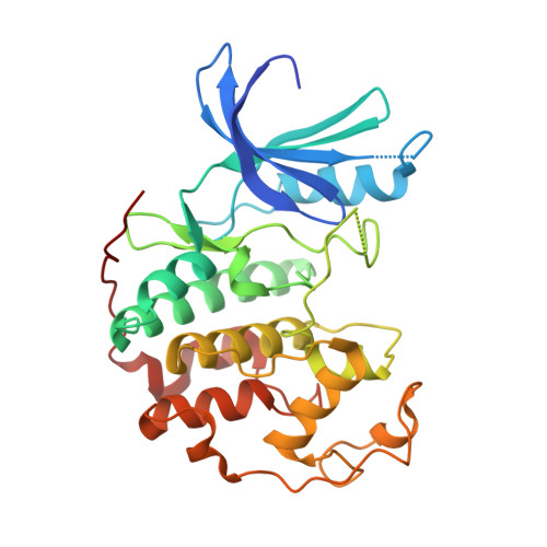
RCSB PDB - 3R8Z: CDK2 in complex with inhibitor RC-1-136

 3R8Z

CDK2 in complex with inhibitor RC-1-136


Experimental Data Snapshot

  • Method: X-RAY DIFFRACTION
  • Resolution: 1.85 Å
  • R-Value Free: 0.251 
  • R-Value Work: 0.207 
  • R-Value Observed: 0.207 

Starting Model: experimental
View more details

wwPDB Validation   3D Report Full Report


Ligand Structure Quality Assessment 

Created with Raphaël 2.3.0Worse 01 BetterLigand structure goodness of fit to experimental dataBest fitted Z63Click on this verticalbar to view details

This is version 1.3 of the entry. See complete history


Literature

Development of highly potent and selective diaminothiazole inhibitors of cyclin-dependent kinases.

Schonbrunn, E.Betzi, S.Alam, R.Martin, M.P.Becker, A.Han, H.Francis, R.Chakrasali, R.Jakkaraj, S.Kazi, A.Sebti, S.M.Cubitt, C.L.Gebhard, A.W.Hazlehurst, L.A.Tash, J.S.Georg, G.I.

(2013) J Med Chem 56: 3768-3782

  • DOI: https://doi.org/10.1021/jm301234k
  • Primary Citation of Related Structures:  
    3QQK, 3QTQ, 3QTR, 3QTS, 3QTU, 3QTW, 3QTX, 3QTZ, 3QU0, 3QXP, 3R8U, 3R8V, 3R8Z, 3R9D, 3R9H, 3R9N, 3R9O, 3RAH, 3RAK, 3RAL, 3RJC, 3RK5, 3RK7, 3RK9, 3RKB, 3RMF, 3RNI, 3RPR, 3RPV, 3RPY, 3RZB, 3S00, 3S0O, 3S1H, 3SQQ, 4GCJ

  • PubMed Abstract: 

    Cyclin-dependent kinases (CDKs) are serine/threonine protein kinases that act as key regulatory elements in cell cycle progression. We describe the development of highly potent diaminothiazole inhibitors of CDK2 (IC50 = 0.0009-0.0015 μM) from a single hit compound with weak inhibitory activity (IC50 = 15 μM), discovered by high-throughput screening. Structure-based design was performed using 35 cocrystal structures of CDK2 liganded with distinct analogues of the parent compound. The profiling of compound 51 against a panel of 339 kinases revealed high selectivity for CDKs, with preference for CDK2 and CDK5 over CDK9, CDK1, CDK4, and CDK6. Compound 51 inhibited the proliferation of 13 out of 15 cancer cell lines with IC50 values between 0.27 and 6.9 μM, which correlated with the complete suppression of retinoblastoma phosphorylation and the onset of apoptosis. Combined, the results demonstrate the potential of this new inhibitors series for further development into CDK-specific chemical probes or therapeutics.


  • Organizational Affiliation

    Drug Discovery Department, H. Lee Moffitt Cancer Center and Research Institute, 12902 Magnolia Drive, Tampa, Florida 33612, USA. ernst.schonbrunn@moffitt.org


Macromolecules
Find similar proteins by:  (by identity cutoff)  |  3D Structure
Entity ID: 1
MoleculeChains Sequence LengthOrganismDetailsImage
Cyclin-dependent kinase 2306Homo sapiensMutation(s): 0 
Gene Names: CDK2
EC: 2.7.11.22
UniProt & NIH Common Fund Data Resources
Find proteins for P24941 (Homo sapiens)
Explore P24941 
Go to UniProtKB:  P24941
PHAROS:  P24941
GTEx:  ENSG00000123374 
Entity Groups  
Sequence Clusters30% Identity50% Identity70% Identity90% Identity95% Identity100% Identity
UniProt GroupP24941
Sequence Annotations
Expand
  • Reference Sequence
Small Molecules
Ligands 1 Unique
IDChains Name / Formula / InChI Key2D Diagram3D Interactions
Z63
Query on Z63

Download Ideal Coordinates CCD File 
B [auth A][4-amino-2-(prop-2-en-1-ylamino)-1,3-thiazol-5-yl](pyridin-4-yl)methanone
C12 H12 N4 O S
JYLDWTJXERPEGH-UHFFFAOYSA-N
Binding Affinity Annotations 
IDSourceBinding Affinity
Z63 PDBBind:  3R8Z IC50: 4.90e+4 (nM) from 1 assay(s)
Experimental Data & Validation

Experimental Data

  • Method: X-RAY DIFFRACTION
  • Resolution: 1.85 Å
  • R-Value Free: 0.251 
  • R-Value Work: 0.207 
  • R-Value Observed: 0.207 
  • Space Group: P 21 21 21
Unit Cell:
Length ( Å )Angle ( ˚ )
a = 53.1α = 90
b = 71.36β = 90
c = 71.28γ = 90
Software Package:
Software NamePurpose
StructureStudiodata collection
CNSrefinement
XDSdata reduction
XDSdata scaling
CNSphasing

Structure Validation

View Full Validation Report


Ligand Structure Quality Assessment 

Created with Raphaël 2.3.0Worse 01 BetterLigand structure goodness of fit to experimental dataBest fitted Z63Click on this verticalbar to view details

Entry History 

Deposition Data

Revision History  (Full details and data files)

  • Version 1.0: 2012-10-31
    Type: Initial release
  • Version 1.1: 2013-05-15
    Changes: Database references
  • Version 1.2: 2013-06-05
    Changes: Database references
  • Version 1.3: 2023-09-13
    Changes: Data collection, Database references, Derived calculations, Refinement description