3RI4

Ets1 cooperative binding to widely separated sites on promoter DNA


Experimental Data Snapshot

  • Method: X-RAY DIFFRACTION
  • Resolution: 3.00 Å
  • R-Value Free: 0.284 
  • R-Value Work: 0.230 
  • R-Value Observed: 0.230 

wwPDB Validation   3D Report Full Report


This is version 1.2 of the entry. See complete history


Literature

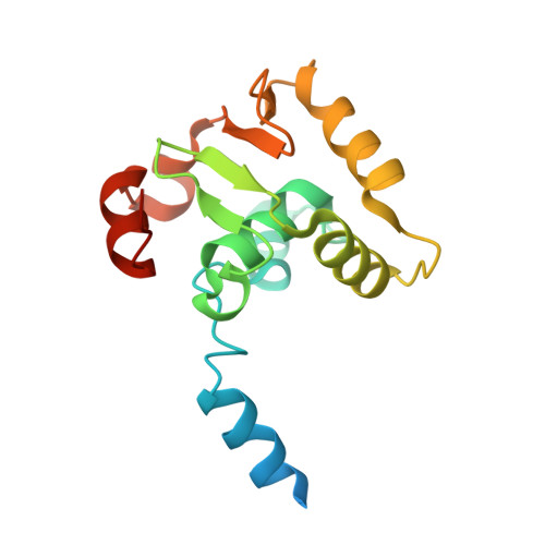
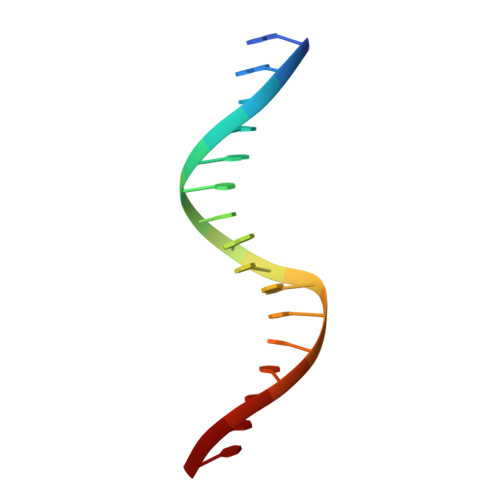
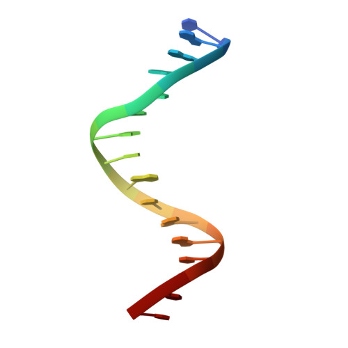
Structural basis of ets1 cooperative binding to widely separated sites on promoter DNA.

Babayeva, N.D.Baranovskaya, O.I.Tahirov, T.H.

(2012) PLoS One 7: e33698-e33698

  • DOI: https://doi.org/10.1371/journal.pone.0033698
  • Primary Citation of Related Structures:  
    3RI4

  • PubMed Abstract: 

    Ets1 is a member of the Ets family of transcription factors. Ets1 is expressed in autoinhibited form and its DNA binding depends on partner proteins bound to adjacent sequences or the relative positioning of a second Ets-binding site (EBS). The autoinhibition of Ets1 is mediated by structural coupling of regions flanking the DNA-binding domain. The NMR structure of Ets1 revealed that the inhibitory regions comprised of helices HI1 and HI2 and H4 are packed together on the Ets domain to form an inhibitory module. The crystal structure of Ets1 unexpectedly revealed a homodimer in which homodimerisation occurs via swapping of HI1 helices. Modeling of DNA binding indicates that the Ets1 dimer can bind to two antiparallel pieces of DNA. To verify this, we crystallized and solved the structure of the complex comprised of Ets1 dimer and two pieces of DNA. DNA binding by Ets1 dimer resulted in formation of additional intermolecular protein•DNA interactions, implying that the complex formation is cooperative.


  • Organizational Affiliation

    Eppley Institute for Research in Cancer and Allied Diseases, University of Nebraska Medical Center, Omaha, Nebraska, United States of America.


Macromolecules

Find similar proteins by:  (by identity cutoff)  |  3D Structure
Entity ID: 1
MoleculeChains Sequence LengthOrganismDetailsImage
Isoform Ets-1 p27 of Protein C-ets-1
A, D
163Homo sapiensMutation(s): 0 
Gene Names: ETS1
UniProt & NIH Common Fund Data Resources
Find proteins for P14921 (Homo sapiens)
Explore P14921 
Go to UniProtKB:  P14921
PHAROS:  P14921
GTEx:  ENSG00000134954 
Entity Groups  
Sequence Clusters30% Identity50% Identity70% Identity90% Identity95% Identity100% Identity
UniProt GroupP14921
Sequence Annotations
Expand
  • Reference Sequence

Find similar nucleic acids by:  Sequence   |   3D Structure  

Entity ID: 2
MoleculeChains LengthOrganismImage
TCR alpha promoter DNA
B, E
16N/A
Sequence Annotations
Expand
  • Reference Sequence

Find similar nucleic acids by:  Sequence   |   3D Structure  

Entity ID: 3
MoleculeChains LengthOrganismImage
TCR alpha promoter DNA
C, F
16N/A
Sequence Annotations
Expand
  • Reference Sequence
Experimental Data & Validation

Experimental Data

  • Method: X-RAY DIFFRACTION
  • Resolution: 3.00 Å
  • R-Value Free: 0.284 
  • R-Value Work: 0.230 
  • R-Value Observed: 0.230 
  • Space Group: P 1 21 1
Unit Cell:
Length ( Å )Angle ( ˚ )
a = 50.177α = 90
b = 98.124β = 109.67
c = 53.577γ = 90
Software Package:
Software NamePurpose
ADSCdata collection
CNSrefinement
HKL-2000data reduction
HKL-2000data scaling
CNSphasing

Structure Validation

View Full Validation Report


Entry History 

Deposition Data

Revision History  (Full details and data files)

  • Version 1.0: 2012-04-04
    Type: Initial release
  • Version 1.1: 2015-12-16
    Changes: Structure summary
  • Version 1.2: 2023-09-13
    Changes: Data collection, Database references, Refinement description, Source and taxonomy, Structure summary