3RIN | pdb_00003rin

p38 kinase crystal structure in complex with small molecule inhibitor


Experimental Data Snapshot

  • Method: X-RAY DIFFRACTION
  • Resolution: 2.20 Å
  • R-Value Free: 
    0.308 (Depositor), 0.311 (DCC) 
  • R-Value Work: 
    0.261 (Depositor), 0.252 (DCC) 
  • R-Value Observed: 
    0.265 (Depositor) 

Starting Model: experimental
View more details

wwPDB Validation 3D Report Full Report

Validation slider image for 3RIN

Ligand Structure Quality Assessment 


This is version 1.1 of the entry. See complete history

Literature

Indolin-2-one p38(alpha) inhibitors I: design, profiling and crystallographic binding mode.

Eastwood, P.Gonzalez, J.Gomez, E.Vidal, B.Caturla, F.Roca, R.Balague, C.Orellana, A.Dominguez, M.

(2011) Bioorg Med Chem Lett 21: 4130-4133

  • DOI: https://doi.org/10.1016/j.bmcl.2011.05.114
  • Primary Citation Related Structures: 
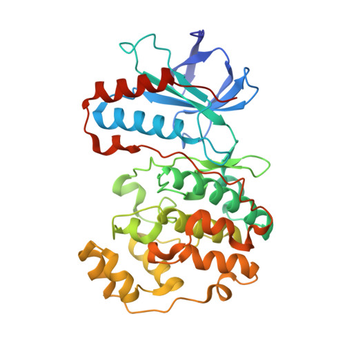
    3RIN

  • PubMed Abstract: 

    The use of structure-based design and molecular modeling led to the discovery of indolin-2-one derivatives as potent and selective p38α inhibitors. The predicted binding mode was confirmed by X-ray crystallography.


  • Organizational Affiliation
    • Almirall Research Center, Almirall S.A., Ctra. Laureà Miró 408, E-08980 St. Feliu de Llobregat, Barcelona, Spain. paul.eastwood@almirall.com

Macromolecule Content 

  • Total Structure Weight: 41.7 kDa 
  • Atom Count: 2,939 
  • Modeled Residue Count: 349 
  • Deposited Residue Count: 360 
  • Unique protein chains: 1

Macromolecules

Find similar proteins by:|  3D Structure
Entity ID: 1
MoleculeChains  Sequence LengthOrganismDetailsImage
Mitogen-activated protein kinase 14360Homo sapiensMutation(s): 0 
Gene Names: CSBPCSBP1CSBP2CSPB1MAPK14MXI2
EC: 2.7.11.24
UniProt & NIH Common Fund Data Resources
Find proteins for Q16539 (Homo sapiens)
Explore Q16539 
Go to UniProtKB:  Q16539
PHAROS:  Q16539
GTEx:  ENSG00000112062 
Entity Groups
Sequence Clusters30% Identity50% Identity70% Identity90% Identity95% Identity100% Identity
UniProt GroupQ16539
Sequence Annotations
Expand
Reference Sequence

Small Molecules

Ligands 1 Unique
IDChains Name / Formula / InChI Key2D Diagram3D Interactions
I2O

Query on I2O


Download:Ideal Coordinates CCD File
B [auth A]N-cyclopropyl-4-methyl-3-(2'-oxo-1',2'-dihydrospiro[cyclopentane-1,3'-indol]-6'-yl)benzamide
C23 H24 N2 O2
WMXJYEDCJIDCNY-UHFFFAOYSA-N

Experimental Data & Validation

Experimental Data

  • Method: X-RAY DIFFRACTION
  • Resolution: 2.20 Å
  • R-Value Free:  0.308 (Depositor), 0.311 (DCC) 
  • R-Value Work:  0.261 (Depositor), 0.252 (DCC) 
  • R-Value Observed: 0.265 (Depositor) 
Space Group: P 21 21 21
Unit Cell:
Length ( Å )Angle ( ˚ )
a = 45.11α = 90
b = 86.62β = 90
c = 123.48γ = 90
Software Package:
Software NamePurpose
REFMACrefinement
PDB_EXTRACTdata extraction
CrystalCleardata collection
CrystalCleardata reduction
CrystalCleardata scaling
MOLREPphasing

Structure Validation

View Full Validation Report


Ligand Structure Quality Assessment 


Entry History 

Deposition Data

Revision History  (Full details and data files)

  • Version 1.0: 2012-02-29
    Type: Initial release
  • Version 1.1: 2023-09-13
    Changes: Data collection, Database references, Derived calculations, Refinement description