3RLD | pdb_00003rld

Crystal structure of the Y7I mutant of human carbonic anhydrase II


Experimental Data Snapshot

  • Method: X-RAY DIFFRACTION
  • Resolution: 1.50 Å
  • R-Value Free: 
    0.211 (Depositor), 0.207 (DCC) 
  • R-Value Work: 
    0.166 (Depositor), 0.176 (DCC) 
  • R-Value Observed: 
    0.166 (Depositor) 

Starting Model: experimental
View more details

wwPDB Validation 3D Report Full Report

Validation slider image for 3RLD

This is version 1.3 of the entry. See complete history

Literature

Kinetic and crystallographic studies of the role of tyrosine 7 in the active site of human carbonic anhydrase II.

Mikulski, R.Avvaru, B.S.Tu, C.Case, N.McKenna, R.Silverman, D.N.

(2011) Arch Biochem Biophys 506: 181-187

  • DOI: https://doi.org/10.1016/j.abb.2010.12.004
  • Primary Citation Related Structures: 
    3RLD

  • PubMed Abstract: 

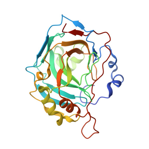
    The rate limiting step in catalysis of bicarbonate dehydration by human carbonic anhydrase II (HCA II) is an intramolecular proton transfer from His64 to the zinc-bound hydroxide. We have examined the role of Tyr7 using site-specific mutagenesis and measuring catalysis by the ¹⁸O exchange method using membrane inlet mass spectrometry. The side chain of Tyr7 in HCA II extends into the active-site cavity about 7 Å from the catalytic zinc atom. Replacement of Tyr7 with eight other amino acids had no effect on the interconversion of bicarbonate and CO₂, but in some cases caused enhancements in the rate constant of proton transfer by nearly 10-fold. The variant Y7I HCA II enhanced intramolecular proton transfer approximately twofold; its structure was determined by X-ray crystallography at 1.5 Å resolution. No changes were observed in the ordered solvent structure in the active-site cavity or in the conformation of the side chain of the proton shuttle His64. However, the first 11 residues of the amino-terminal chain in Y7I HCA II assumed an alternate conformation compared with the wild type. Differential scanning calorimetry showed variants at position 7 had a melting temperature approximately 8 °C lower than that of the wild type.


  • Organizational Affiliation
    • Department of Pharmacology, University of Florida, Gainesville, FL 32610, USA.

Macromolecule Content 

  • Total Structure Weight: 29.59 kDa 
  • Atom Count: 2,261 
  • Modeled Residue Count: 257 
  • Deposited Residue Count: 260 
  • Unique protein chains: 1

Macromolecules

Find similar proteins by:|  3D Structure
Entity ID: 1
MoleculeChains  Sequence LengthOrganismDetailsImage
Carbonic anhydrase 2260Homo sapiensMutation(s): 1 
Gene Names: CA2
EC: 4.2.1.1 (PDB Primary Data), 4.2.1.69 (UniProt)
UniProt & NIH Common Fund Data Resources
Find proteins for P00918 (Homo sapiens)
Explore P00918 
Go to UniProtKB:  P00918
PHAROS:  P00918
GTEx:  ENSG00000104267 
Entity Groups
Sequence Clusters30% Identity50% Identity70% Identity90% Identity95% Identity100% Identity
UniProt GroupP00918
Sequence Annotations
Expand
Reference Sequence

Experimental Data & Validation

Experimental Data

  • Method: X-RAY DIFFRACTION
  • Resolution: 1.50 Å
  • R-Value Free:  0.211 (Depositor), 0.207 (DCC) 
  • R-Value Work:  0.166 (Depositor), 0.176 (DCC) 
  • R-Value Observed: 0.166 (Depositor) 
Space Group: P 1 21 1
Unit Cell:
Length ( Å )Angle ( ˚ )
a = 42.789α = 90
b = 41.598β = 104.83
c = 73.195γ = 90
Software Package:
Software NamePurpose
DENZOdata reduction
SCALEPACKdata scaling
SHELXrefinement
PDB_EXTRACTdata extraction
CrystalCleardata collection
HKL-2000data reduction
CNSphasing
SHELXLrefinement

Structure Validation

View Full Validation Report


Entry History 

Deposition Data

Revision History  (Full details and data files)

  • Version 1.0: 2012-02-08
    Type: Initial release
  • Version 1.1: 2012-03-14
    Changes: Other
  • Version 1.2: 2017-11-08
    Changes: Refinement description
  • Version 1.3: 2023-09-13
    Changes: Data collection, Database references, Derived calculations, Refinement description