3RWE

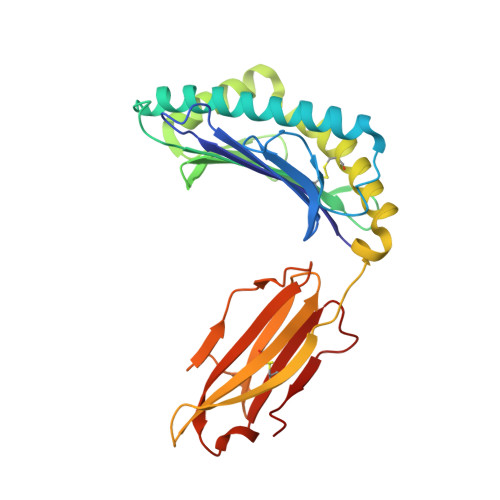
rhesus macaque MHC class I molecule Mamu-B*17-FW9


Experimental Data Snapshot

  • Method: X-RAY DIFFRACTION
  • Resolution: 2.40 Å
  • R-Value Free: 0.251 
  • R-Value Work: 0.203 
  • R-Value Observed: 0.206 

Starting Model: experimental
View more details

wwPDB Validation   3D Report Full Report


This is version 1.2 of the entry. See complete history


Literature

Structural basis of diverse peptide accommodation by the rhesus macaque MHC class I molecule Mamu-B*17: insights into immune protection from simian immunodeficiency virus

Wu, Y.Gao, F.Liu, J.Qi, J.X.Gostick, E.Price, D.A.Gao, G.F.

(2011) J Immunol 187: 6382-6392

  • DOI: https://doi.org/10.4049/jimmunol.1101726
  • Primary Citation of Related Structures:  
    3RWC, 3RWD, 3RWE, 3RWF, 3RWG, 3RWH, 3RWI, 3RWJ

  • PubMed Abstract: 

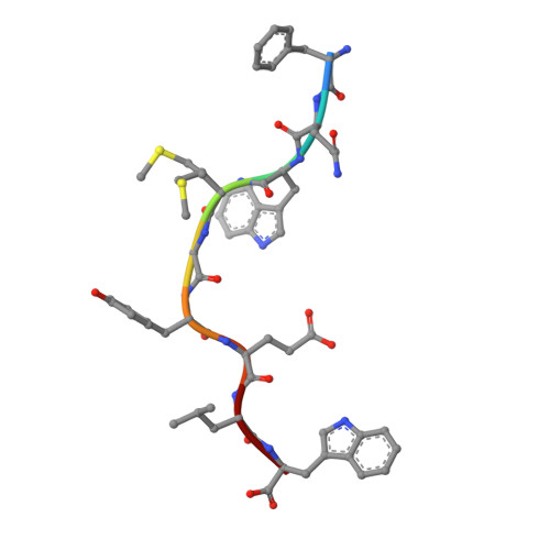
    The MHC class I molecule Mamu-B*17 has been associated with elite control of SIV infection in rhesus macaques, akin to the protective effects described for HLA-B*57 in HIV-infected individuals. In this study, we determined the crystal structures of Mamu-B*17 in complex with eight different peptides corresponding to immunodominant SIV(mac)239-derived CD8(+) T cell epitopes: HW8 (HLEVQGYW), GW10 (GSHLEVQGYW), MW9 (MHPAQTSQW), QW9 (QTSQWDDPW), FW9 (FQWMGYELW), MF8 (MRHVLEPF), IW9 (IRYPKTFGW), and IW11 (IRYPKTFGWLW). The structures reveal that not only P2, but also P1 and P3, can be used as N-terminal anchor residues by Mamu-B*17-restricted peptides. Moreover, the N-terminal anchor residues exhibit a broad chemical specificity, encompassing basic (H and R), bulky polar aliphatic (Q), and small (T) residues. In contrast, Mamu-B*17 exhibits a very narrow preference for aromatic residues (W and F) at the C terminus, similar to that displayed by HLA-B*57. Flexibility within the whole peptide-binding groove contributes to the accommodation of these diverse peptides, which adopt distinct conformations. Furthermore, the unusually large pocket D enables compensation from other peptide residues if P3 is occupied by an amino acid with a small side chain. In addition, residues located at likely TCR contact regions present highly flexible conformations, which may impact TCR repertoire profiles. These findings provide novel insights into the structural basis of diverse peptide accommodation by Mamu-B*17 and highlight unique atomic features that might contribute to the protective effect of this MHC I molecule in SIV-infected rhesus macaques.


  • Organizational Affiliation

    CAS Key Laboratory of Pathogenic Microbiology and Immunology, Institute of Microbiology, Chinese Academy of Sciences, Beijing 100101, The People's Republic of China.


Macromolecules
Find similar proteins by:  (by identity cutoff)  |  3D Structure
Entity ID: 1
MoleculeChains Sequence LengthOrganismDetailsImage
Major histocompatibility complex class I276Macaca mulattaMutation(s): 0 
Gene Names: MHCI-B
UniProt
Find proteins for Q9GJ77 (Macaca mulatta)
Explore Q9GJ77 
Go to UniProtKB:  Q9GJ77
Entity Groups  
Sequence Clusters30% Identity50% Identity70% Identity90% Identity95% Identity100% Identity
UniProt GroupQ9GJ77
Sequence Annotations
Expand
  • Reference Sequence
Find similar proteins by:  (by identity cutoff)  |  3D Structure
Entity ID: 2
MoleculeChains Sequence LengthOrganismDetailsImage
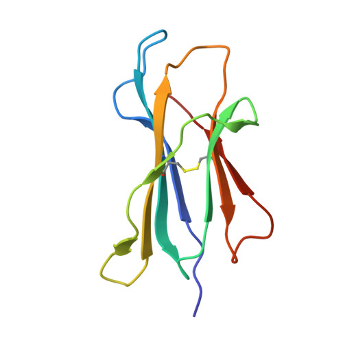
Beta-2-microglobulin99Macaca mulattaMutation(s): 0 
Gene Names: B2M
UniProt
Find proteins for Q6V7J5 (Macaca mulatta)
Explore Q6V7J5 
Go to UniProtKB:  Q6V7J5
Entity Groups  
Sequence Clusters30% Identity50% Identity70% Identity90% Identity95% Identity100% Identity
UniProt GroupQ6V7J5
Sequence Annotations
Expand
  • Reference Sequence

Find similar proteins by:  Sequence   |   3D Structure  

Entity ID: 3
MoleculeChains Sequence LengthOrganismDetailsImage
Pol FW9 peptide from Pol protein9Simian immunodeficiency virusMutation(s): 0 
UniProt
Find proteins for Q5QGH9 (Simian immunodeficiency virus)
Explore Q5QGH9 
Go to UniProtKB:  Q5QGH9
Entity Groups  
UniProt GroupQ5QGH9
Sequence Annotations
Expand
  • Reference Sequence
Experimental Data & Validation

Experimental Data

  • Method: X-RAY DIFFRACTION
  • Resolution: 2.40 Å
  • R-Value Free: 0.251 
  • R-Value Work: 0.203 
  • R-Value Observed: 0.206 
  • Space Group: P 1 2 1
Unit Cell:
Length ( Å )Angle ( ˚ )
a = 68.808α = 90
b = 45.29β = 96.11
c = 81.982γ = 90
Software Package:
Software NamePurpose
PHENIXrefinement

Structure Validation

View Full Validation Report


Entry History 

Deposition Data

Revision History  (Full details and data files)

  • Version 1.0: 2012-03-21
    Type: Initial release
  • Version 1.1: 2023-11-01
    Changes: Data collection, Database references, Derived calculations, Refinement description
  • Version 1.2: 2024-10-30
    Changes: Structure summary