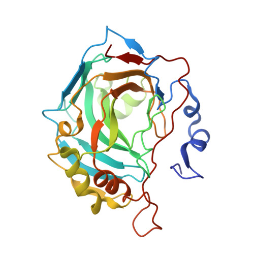
Fluoroalkyl and alkyl chains have similar hydrophobicities in binding to the "hydrophobic wall" of carbonic anhydrase.
Mecinovic, J., Snyder, P.W., Mirica, K.A., Bai, S., Mack, E.T., Kwant, R.L., Moustakas, D.T., Heroux, A., Whitesides, G.M.(2011) J Am Chem Soc 133: 14017-14026
- PubMed: 21790183
- DOI: https://doi.org/10.1021/ja2045293
- Primary Citation of Related Structures:
3RYJ, 3RYV, 3RYX, 3RYY, 3RYZ, 3RZ0, 3RZ1, 3RZ5, 3RZ7, 3RZ8 - PubMed Abstract:
The hydrophobic effect, the free-energetically favorable association of nonpolar solutes in water, makes a dominant contribution to binding of many systems of ligands and proteins. The objective of this study was to examine the hydrophobic effect in biomolecular recognition using two chemically different but structurally similar hydrophobic groups, aliphatic hydrocarbons and aliphatic fluorocarbons, and to determine whether the hydrophobicity of the two groups could be distinguished by thermodynamic and biostructural analysis. This paper uses isothermal titration calorimetry (ITC) to examine the thermodynamics of binding of benzenesulfonamides substituted in the para position with alkyl and fluoroalkyl chains (H(2)NSO(2)C(6)H(4)-CONHCH(2)(CX(2))(n)CX(3), n = 0-4, X = H, F) to human carbonic anhydrase II (HCA II). Both alkyl and fluoroalkyl substituents contribute favorably to the enthalpy and the entropy of binding; these contributions increase as the length of chain of the hydrophobic substituent increases. Crystallography of the protein-ligand complexes indicates that the benzenesulfonamide groups of all ligands examined bind with similar geometry, that the tail groups associate with the hydrophobic wall of HCA II (which is made up of the side chains of residues Phe131, Val135, Pro202, and Leu204), and that the structure of the protein is indistinguishable for all but one of the complexes (the longest member of the fluoroalkyl series). Analysis of the thermodynamics of binding as a function of structure is compatible with the hypothesis that hydrophobic binding of both alkyl and fluoroalkyl chains to hydrophobic surface of carbonic anhydrase is due primarily to the release of nonoptimally hydrogen-bonded water molecules that hydrate the binding cavity (including the hydrophobic wall) of HCA II and to the release of water molecules that surround the hydrophobic chain of the ligands. This study defines the balance of enthalpic and entropic contributions to the hydrophobic effect in this representative system of protein and ligand: hydrophobic interactions, here, seem to comprise approximately equal contributions from enthalpy (plausibly from strengthening networks of hydrogen bonds among molecules of water) and entropy (from release of water from configurationally restricted positions).
- Department of Chemistry and Chemical Biology, Harvard University, Cambridge, Massachusetts 02138, United States.
Organizational Affiliation:


