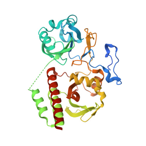
3S7Q

Crystal Structure of a Monomeric Infrared Fluorescent Deinococcus radiodurans Bacteriophytochrome chromophore binding domain


Experimental Data Snapshot

  • Method: X-RAY DIFFRACTION
  • Resolution: 1.75 Å
  • R-Value Free: 0.217 
  • R-Value Work: 0.187 
  • R-Value Observed: 0.189 

Starting Model: experimental
View more details

wwPDB Validation   3D Report Full Report


Ligand Structure Quality Assessment 


This is version 1.3 of the entry. See complete history


Literature

Structure-guided engineering enhances a phytochrome-based infrared fluorescent protein.

Auldridge, M.E.Satyshur, K.A.Anstrom, D.M.Forest, K.T.

(2012) J Biol Chem 287: 7000-7009

  • DOI: https://doi.org/10.1074/jbc.M111.295121
  • Primary Citation of Related Structures:  
    3S7N, 3S7O, 3S7P, 3S7Q

  • PubMed Abstract: 

    Phytochrome is a multidomain dimeric red light photoreceptor that utilizes a chromophore-binding domain (CBD), a PHY domain, and an output module to induce cellular changes in response to light. A promising biotechnology tool emerged when a structure-based substitution at Asp-207 was shown to be an infrared fluorophore that uses a biologically available tetrapyrrole chromophore. We report multiple crystal structures of this D207H variant of the Deinococcus radiodurans CBD, in which His-207 is observed to form a hydrogen bond with either the tetrapyrrole A-ring oxygen or the Tyr-263 hydroxyl. Based on the implications of this duality for fluorescence properties, Y263F was introduced and shown to have stronger fluorescence than the original D207H template. Our structures are consistent with the model that the Y263F change prevents a red light-induced far-red light absorbing phytochrome chromophore configuration. With the goal of decreasing size and thereby facilitating use as a fluorescent tag in vivo, we also engineered a monomeric form of the CBD. Unexpectedly, photoconversion was observed in the monomer despite the lack of a PHY domain. This observation underscores an interplay between dimerization and the photochemical properties of phytochrome and suggests that the monomeric CBD could be used for further studies of the photocycle. The D207H substitution on its own in the monomer did not result in fluorescence, whereas Y263F did. Combined, the D207H and Y263F substitutions in the monomeric CBD lead to the brightest of our variants, designated Wisconsin infrared phytofluor (Wi-Phy).


  • Organizational Affiliation

    Department of Bacteriology, University of Wisconsin-Madison, Madison, Wisconsin 53706, USA.


Macromolecules
Find similar proteins by:  (by identity cutoff)  |  3D Structure
Entity ID: 1
MoleculeChains Sequence LengthOrganismDetailsImage
Bacteriophytochrome343Deinococcus radioduransMutation(s): 5 
Gene Names: bphPDR_A0050
EC: 2.7.13.3
UniProt
Find proteins for Q9RZA4 (Deinococcus radiodurans (strain ATCC 13939 / DSM 20539 / JCM 16871 / CCUG 27074 / LMG 4051 / NBRC 15346 / NCIMB 9279 / VKM B-1422 / R1))
Explore Q9RZA4 
Go to UniProtKB:  Q9RZA4
Entity Groups  
Sequence Clusters30% Identity50% Identity70% Identity90% Identity95% Identity100% Identity
UniProt GroupQ9RZA4
Sequence Annotations
Expand
  • Reference Sequence
Small Molecules
Ligands 4 Unique
IDChains Name / Formula / InChI Key2D Diagram3D Interactions
LBW
Query on LBW

Download Ideal Coordinates CCD File 
C [auth A]3-[2-[(Z)-[5-[(Z)-(4-ethenyl-3-methyl-5-oxidanylidene-pyrrol-2-ylidene)methyl]-3-(3-hydroxy-3-oxopropyl)-4-methyl-pyrrol-1-ium-2-ylidene]methyl]-5-[(Z)-[(3E,4R)-3-ethylidene-4-methyl-5-oxidanylidene-pyrrolidin-2-ylidene]methyl]-4-methyl-1H-pyrrol-3-yl]propanoic acid
C33 H37 N4 O6
DKMLMZVDTGOEGU-UAWLBFNISA-O
LBV
Query on LBV

Download Ideal Coordinates CCD File 
B [auth A]3-[2-[(Z)-[3-(2-carboxyethyl)-5-[(Z)-(4-ethenyl-3-methyl-5-oxidanylidene-pyrrol-2-ylidene)methyl]-4-methyl-pyrrol-1-ium -2-ylidene]methyl]-5-[(Z)-[(3E)-3-ethylidene-4-methyl-5-oxidanylidene-pyrrolidin-2-ylidene]methyl]-4-methyl-1H-pyrrol-3- yl]propanoic acid
C33 H37 N4 O6
DKMLMZVDTGOEGU-ISEYCTJISA-O
PO4
Query on PO4

Download Ideal Coordinates CCD File 
F [auth A]PHOSPHATE ION
O4 P
NBIIXXVUZAFLBC-UHFFFAOYSA-K
GOL
Query on GOL

Download Ideal Coordinates CCD File 
D [auth A],
E [auth A]
GLYCEROL
C3 H8 O3
PEDCQBHIVMGVHV-UHFFFAOYSA-N
Experimental Data & Validation

Experimental Data

  • Method: X-RAY DIFFRACTION
  • Resolution: 1.75 Å
  • R-Value Free: 0.217 
  • R-Value Work: 0.187 
  • R-Value Observed: 0.189 
  • Space Group: C 1 2 1
Unit Cell:
Length ( Å )Angle ( ˚ )
a = 95.129α = 90
b = 55.072β = 92.23
c = 69.969γ = 90
Software Package:
Software NamePurpose
PHASERphasing
REFMACrefinement
PDB_EXTRACTdata extraction
MAR345dtbdata collection
HKL-2000data reduction
HKL-2000data scaling

Structure Validation

View Full Validation Report


Ligand Structure Quality Assessment 


Entry History 

Deposition Data

Revision History  (Full details and data files)

  • Version 1.0: 2012-01-11
    Type: Initial release
  • Version 1.1: 2012-11-28
    Changes: Database references
  • Version 1.2: 2023-09-13
    Changes: Data collection, Database references, Derived calculations, Refinement description, Structure summary
  • Version 1.3: 2024-10-16
    Changes: Structure summary