3S8E | pdb_00003s8e

Phosphorylation regulates assembly of the caspase-6 substrate-binding groove


Experimental Data Snapshot

  • Method: X-RAY DIFFRACTION
  • Resolution: 2.88 Å
  • R-Value Free: 
    0.256 (Depositor), 0.250 (DCC) 
  • R-Value Work: 
    0.216 (Depositor), 0.211 (DCC) 
  • R-Value Observed: 
    0.218 (Depositor) 

Starting Model: experimental
View more details

wwPDB Validation 3D Report Full Report

Validation slider image for 3S8E

This is version 1.2 of the entry. See complete history

Literature

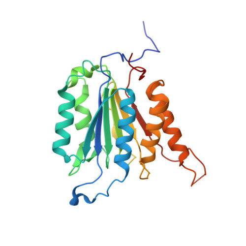
Phosphorylation regulates assembly of the caspase-6 substrate-binding groove.

Velazquez-Delgado, E.M.Hardy, J.A.

(2012) Structure 20: 742-751

  • DOI: https://doi.org/10.1016/j.str.2012.02.003
  • Primary Citation Related Structures: 
    3S8E

  • PubMed Abstract: 

    Caspases, a family of apoptotic proteases, are increasingly recognized as being extensively phosphorylated, usually leading to inactivation. To date, no structural mechanism for phosphorylation-based caspase inactivation is available, although this information may be key to achieving caspase-specific inhibition. Caspase-6 has recently been implicated in neurodegenerative conditions including Huntington's and Alzheimer's diseases. A full understanding of caspase-6 regulation is crucial to caspase-6-specific inhibition. Caspase-6 is phosphorylated by ARK5 kinase at serine 257 leading to suppression of cell death via caspase-6 inhibition. Our structure of the fully inactive phosphomimetic S257D reveals that phosphorylation results in a steric clash with P201 in the L2' loop. Removal of the proline side chain alleviates the clash resulting in nearly wild-type activity levels. This phosphomimetic-mediated steric clash causes misalignment of the substrate-binding groove, preventing substrate binding. Substrate-binding loop misalignment appears to be a widely used regulatory strategy among caspases and may present a new paradigm for caspase-specific control.


  • Organizational Affiliation
    • Department of Chemistry, University of Massachusetts Amherst, Amherst, MA 01003, USA.

Macromolecule Content 

  • Total Structure Weight: 255.77 kDa 
  • Atom Count: 13,881 
  • Modeled Residue Count: 1,747 
  • Deposited Residue Count: 2,216 
  • Unique protein chains: 1

Macromolecules

Find similar proteins by:|  3D Structure
Entity ID: 1
MoleculeChains  Sequence LengthOrganismDetailsImage
Caspase-6
A, B, C, D, E
A, B, C, D, E, F, G, H
277Homo sapiensMutation(s): 1 
Gene Names: CASP6MCH2
EC: 3.4.22.59
UniProt & NIH Common Fund Data Resources
Find proteins for P55212 (Homo sapiens)
Explore P55212 
Go to UniProtKB:  P55212
PHAROS:  P55212
GTEx:  ENSG00000138794 
Entity Groups
Sequence Clusters30% Identity50% Identity70% Identity90% Identity95% Identity100% Identity
UniProt GroupP55212
Sequence Annotations
Expand
Reference Sequence

Experimental Data & Validation

Experimental Data

  • Method: X-RAY DIFFRACTION
  • Resolution: 2.88 Å
  • R-Value Free:  0.256 (Depositor), 0.250 (DCC) 
  • R-Value Work:  0.216 (Depositor), 0.211 (DCC) 
  • R-Value Observed: 0.218 (Depositor) 
Space Group: P 1 21 1
Unit Cell:
Length ( Å )Angle ( ˚ )
a = 81.671α = 90
b = 163.662β = 94.17
c = 89.015γ = 90
Software Package:
Software NamePurpose
CrystalCleardata collection
PHASERphasing
REFMACrefinement
HKL-2000data reduction
SCALEPACKdata scaling

Structure Validation

View Full Validation Report


Entry History 

Deposition Data

Revision History  (Full details and data files)

  • Version 1.0: 2012-04-11
    Type: Initial release
  • Version 1.1: 2013-07-10
    Changes: Database references
  • Version 1.2: 2023-11-01
    Changes: Data collection, Database references, Refinement description