An intrinsically labile alpha-helix abutting the BCL9-binding site of beta-catenin is required for its inhibition by carnosic acid.
de la Roche, M., Rutherford, T.J., Gupta, D., Veprintsev, D.B., Saxty, B., Freund, S.M., Bienz, M.(2012) Nat Commun 3: 680-680
- PubMed: 22353711 Search on PubMedSearch on PubMed Central
- DOI: https://doi.org/10.1038/ncomms1680
- Primary Citation Related Structures:
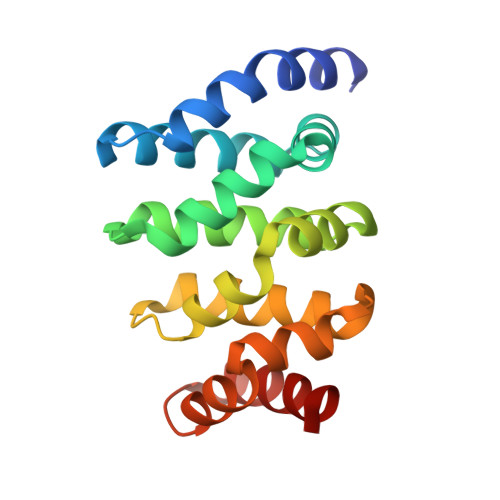
3SL9, 3SLA - PubMed Abstract:
Wnt/β-catenin signalling controls development and tissue homeostasis. Moreover, activated β-catenin can be oncogenic and, notably, drives colorectal cancer. Inhibiting oncogenic β-catenin has proven a formidable challenge. Here we design a screen for small-molecule inhibitors of β-catenin's binding to its cofactor BCL9, and discover five related natural compounds, including carnosic acid from rosemary, which attenuates transcriptional β-catenin outputs in colorectal cancer cells. Evidence from NMR and analytical ultracentrifugation demonstrates that the carnosic acid response requires an intrinsically labile α-helix (H1) amino-terminally abutting the BCL9-binding site in β-catenin. Similarly, in colorectal cancer cells with hyperactive β-catenin signalling, carnosic acid targets predominantly the transcriptionally active ('oncogenic') form of β-catenin for proteasomal degradation in an H1-dependent manner. Hence, H1 is an 'Achilles' Heel' of β-catenin, which can be exploited for destabilization of oncogenic β-catenin by small molecules, providing proof-of-principle for a new strategy for developing direct inhibitors of oncogenic β-catenin.
- MRC Laboratory of Molecular Biology, Hills Road, Cambridge CB2 0QH, UK.
Organizational Affiliation: