3T8O

Rhodopsin kinase (GRK1) L166K mutant at 2.5A resolution


Experimental Data Snapshot

  • Method: X-RAY DIFFRACTION
  • Resolution: 2.50 Å
  • R-Value Free: 
    0.245 (Depositor), 0.240 (DCC) 
  • R-Value Work: 
    0.207 (Depositor), 0.200 (DCC) 
  • R-Value Observed: 
    0.209 (Depositor) 

wwPDB Validation   3D Report Full Report


Ligand Structure Quality Assessment 

Created with Raphaël 2.3.0Worse 01 BetterLigand structure goodness of fit to experimental dataBest fitted ATPClick on this verticalbar to view details

This is version 1.3 of the entry. See complete history


Literature

Structure of a monomeric variant of rhodopsin kinase at 2.5 A resolution.

Tesmer, J.J.Nance, M.R.Singh, P.Lee, H.

(2012) Acta Crystallogr Sect F Struct Biol Cryst Commun 68: 622-625

  • DOI: https://doi.org/10.1107/S1744309112017435
  • Primary Citation of Related Structures:  
    3T8O

  • PubMed Abstract: 

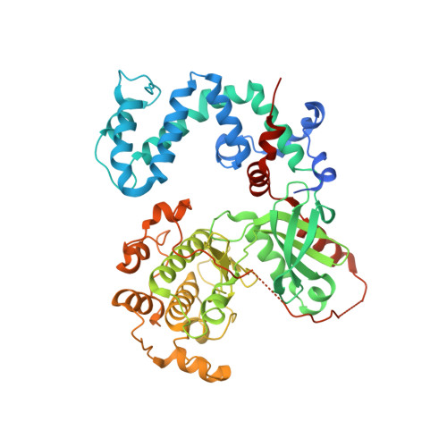
    G protein-coupled receptor kinase 1 (GRK1 or rhodopsin kinase) phosphorylates activated rhodopsin and initiates a cascade of events that results in the termination of phototransduction by the receptor. Although GRK1 seems to be a monomer in solution, seven prior crystal structures of GRK1 revealed a similar domain-swapped dimer interface involving the C-terminus of the enzyme. The influence of this interface on the overall conformation of GRK1 is not known. To address this question, the crystalline dimer interface was disrupted with a L166K mutation and the structure of GRK1-L166K was determined in complex with Mg(2+) · ATP to 2.5 Å resolution. GRK1-L166K crystallized in a novel space group as a monomer and exhibited little overall conformational difference from prior structures of GRK1, although the C-terminal domain-swapped region had reorganized owing to loss of the dimer interface.


  • Organizational Affiliation

    Life Sciences Institute and the Department of Pharmacology, University of Michigan, 210 Washtenaw Avenue, Ann Arbor, MI 48109-2216, USA. tesmerjj@umich.edu


Macromolecules
Find similar proteins by:  (by identity cutoff)  |  3D Structure
Entity ID: 1
MoleculeChains Sequence LengthOrganismDetailsImage
Rhodopsin kinase543Bos taurusMutation(s): 1 
Gene Names: GRK1RHOK
EC: 2.7.11.14
UniProt
Find proteins for P28327 (Bos taurus)
Explore P28327 
Go to UniProtKB:  P28327
Entity Groups  
Sequence Clusters30% Identity50% Identity70% Identity90% Identity95% Identity100% Identity
UniProt GroupP28327
Sequence Annotations
Expand
  • Reference Sequence
Experimental Data & Validation

Experimental Data

  • Method: X-RAY DIFFRACTION
  • Resolution: 2.50 Å
  • R-Value Free:  0.245 (Depositor), 0.240 (DCC) 
  • R-Value Work:  0.207 (Depositor), 0.200 (DCC) 
  • R-Value Observed: 0.209 (Depositor) 
Space Group: I 2 2 2
Unit Cell:
Length ( Å )Angle ( ˚ )
a = 55.541α = 90
b = 149.808β = 90
c = 190.879γ = 90
Software Package:
Software NamePurpose
DENZOdata reduction
SCALEPACKdata scaling
REFMACrefinement
PDB_EXTRACTdata extraction

Structure Validation

View Full Validation Report


Ligand Structure Quality Assessment 

Created with Raphaël 2.3.0Worse 01 BetterLigand structure goodness of fit to experimental dataBest fitted ATPClick on this verticalbar to view details

Entry History 

Deposition Data

Revision History  (Full details and data files)

  • Version 1.0: 2012-06-06
    Type: Initial release
  • Version 1.1: 2013-01-23
    Changes: Database references
  • Version 1.2: 2017-11-08
    Changes: Refinement description
  • Version 1.3: 2024-02-28
    Changes: Data collection, Database references, Derived calculations