3UFT | pdb_00003uft

Structure of rat nitric oxide synthase heme domain in complex with 6-(((3R,4R)-4-(4-(3-chloro-5-fluorophenoxy)butoxy)pyrrolidin-3-yl)methyl)-4-methylpyridin-2-amine


Experimental Data Snapshot

  • Method: X-RAY DIFFRACTION
  • Resolution: 2.08 Å
  • R-Value Free: 
    0.260 (Depositor), 0.274 (DCC) 
  • R-Value Work: 
    0.206 (Depositor), 0.233 (DCC) 
  • R-Value Observed: 
    0.208 (Depositor) 

Starting Model: experimental
View more details

wwPDB Validation 3D Report Full Report

Validation slider image for 3UFT

Ligand Structure Quality Assessment 


This is version 1.3 of the entry. See complete history

Literature

Selective monocationic inhibitors of neuronal nitric oxide synthase. Binding mode insights from molecular dynamics simulations.

Huang, H.Ji, H.Li, H.Jing, Q.Labby, K.J.Martasek, P.Roman, L.J.Poulos, T.L.Silverman, R.B.

(2012) J Am Chem Soc 134: 11559-11572

  • DOI: https://doi.org/10.1021/ja302269r
  • Primary Citation Related Structures: 
    3UFO, 3UFP, 3UFQ, 3UFR, 3UFS, 3UFT, 3UFU, 3UFV, 3UFW, 4EUX

  • PubMed Abstract: 

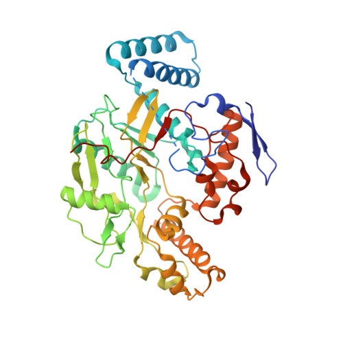
    The reduction of pathophysiologic levels of nitric oxide through inhibition of neuronal nitric oxide synthase (nNOS) has the potential to be therapeutically beneficial in various neurodegenerative diseases. We have developed a series of pyrrolidine-based nNOS inhibitors that exhibit excellent potencies and isoform selectivities (J. Am. Chem. Soc. 2010, 132, 5437). However, there are still important challenges, such as how to decrease the multiple positive charges derived from basic amino groups, which contribute to poor bioavailability, without losing potency and/or selectivity. Here we present an interdisciplinary study combining molecular docking, crystallography, molecular dynamics simulations, synthesis, and enzymology to explore potential pharmacophoric features of nNOS inhibitors and to design potent and selective monocationic nNOS inhibitors. The simulation results indicate that different hydrogen bond patterns, electrostatic interactions, hydrophobic interactions, and a water molecule bridge are key factors for stabilizing ligands and controlling ligand orientation. We find that a heteroatom in the aromatic head or linker chain of the ligand provides additional stability and blocks the substrate binding pocket. Finally, the computational insights are experimentally validated with double-headed pyridine analogues. The compounds reported here are among the most potent and selective monocationic pyrrolidine-based nNOS inhibitors reported to date, and 10 shows improved membrane permeability.


  • Organizational Affiliation
    • Department of Chemistry, Chemistry of Life Processes Institute, amd Center for Molecular Innovation and Drug Discovery, Northwestern University, 2145 Sheridan Road, Evanston, Illinois 60208-3113, USA.

Macromolecule Content 

  • Total Structure Weight: 100.34 kDa 
  • Atom Count: 7,013 
  • Modeled Residue Count: 818 
  • Deposited Residue Count: 844 
  • Unique protein chains: 1

Macromolecules

Find similar proteins by:|  3D Structure
Entity ID: 1
MoleculeChains  Sequence LengthOrganismDetailsImage
Nitric oxide synthase, brain
A, B
422Rattus norvegicusMutation(s): 0 
Gene Names: Nos1Bnos
EC: 1.14.13.39
UniProt
Find proteins for P29476 (Rattus norvegicus)
Explore P29476 
Go to UniProtKB:  P29476
Entity Groups
Sequence Clusters30% Identity50% Identity70% Identity90% Identity95% Identity100% Identity
UniProt GroupP29476
Sequence Annotations
Expand
Reference Sequence

Small Molecules

Ligands 5 Unique
IDChains Name / Formula / InChI Key2D Diagram3D Interactions
HEM

Query on HEM


Download:Ideal Coordinates CCD File
C [auth A],
H [auth B]
PROTOPORPHYRIN IX CONTAINING FE
C34 H32 Fe N4 O4
KABFMIBPWCXCRK-RGGAHWMASA-L
HW7

Query on HW7


Download:Ideal Coordinates CCD File
E [auth A],
J [auth B]
6-({(3R,4R)-4-[4-(3-chloro-5-fluorophenoxy)butoxy]pyrrolidin-3-yl}methyl)-4-methylpyridin-2-amine
C21 H27 Cl F N3 O2
AXHBVFDWXKWFTE-QRWLVFNGSA-N
H4B

Query on H4B


Download:Ideal Coordinates CCD File
D [auth A],
I [auth B]
5,6,7,8-TETRAHYDROBIOPTERIN
C9 H15 N5 O3
FNKQXYHWGSIFBK-RPDRRWSUSA-N
ZN

Query on ZN


Download:Ideal Coordinates CCD File
G [auth A]ZINC ION
Zn
PTFCDOFLOPIGGS-UHFFFAOYSA-N
ACT

Query on ACT


Download:Ideal Coordinates CCD File
F [auth A],
K [auth B]
ACETATE ION
C2 H3 O2
QTBSBXVTEAMEQO-UHFFFAOYSA-M

Experimental Data & Validation

Experimental Data

  • Method: X-RAY DIFFRACTION
  • Resolution: 2.08 Å
  • R-Value Free:  0.260 (Depositor), 0.274 (DCC) 
  • R-Value Work:  0.206 (Depositor), 0.233 (DCC) 
  • R-Value Observed: 0.208 (Depositor) 
Space Group: P 21 21 21
Unit Cell:
Length ( Å )Angle ( ˚ )
a = 51.845α = 90
b = 111.224β = 90
c = 164.111γ = 90
Software Package:
Software NamePurpose
Blu-Icedata collection
REFMACrefinement
HKL-2000data reduction
SCALEPACKdata scaling
REFMACphasing

Structure Validation

View Full Validation Report


Ligand Structure Quality Assessment 


Entry History 

Deposition Data

Revision History  (Full details and data files)

  • Version 1.0: 2012-07-11
    Type: Initial release
  • Version 1.1: 2012-08-01
    Changes: Database references
  • Version 1.2: 2017-11-08
    Changes: Refinement description
  • Version 1.3: 2023-09-13
    Changes: Data collection, Database references, Derived calculations, Refinement description