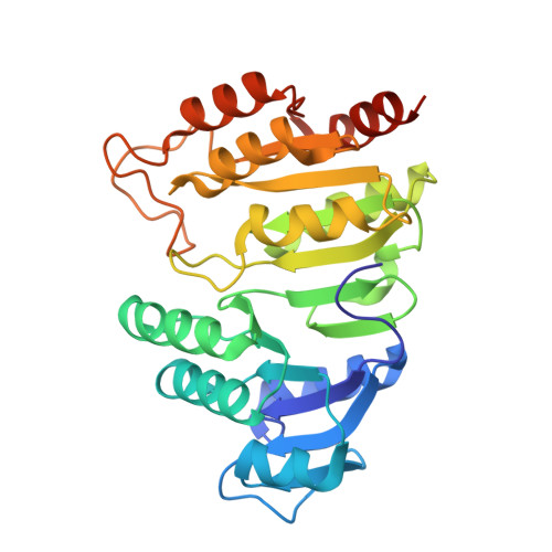
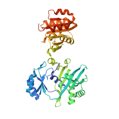
3UFX

Thermus aquaticus succinyl-CoA synthetase in complex with GDP-Mn2+


Experimental Data Snapshot

  • Method: X-RAY DIFFRACTION
  • Resolution: 2.35 Å
  • R-Value Free: 0.245 
  • R-Value Work: 0.204 
  • R-Value Observed: 0.206 

Starting Models: experimental
View more details

wwPDB Validation   3D Report Full Report


Ligand Structure Quality Assessment 


This is version 1.3 of the entry. See complete history


Literature

Biochemical and structural characterization of the GTP-preferring succinyl-CoA synthetase from Thermus aquaticus.

Joyce, M.A.Hayakawa, K.Wolodko, W.T.Fraser, M.E.

(2012) Acta Crystallogr D Biol Crystallogr 68: 751-762

  • DOI: https://doi.org/10.1107/S0907444912010852
  • Primary Citation of Related Structures:  
    3UFX

  • PubMed Abstract: 

    Succinyl-CoA synthetase (SCS) from Thermus aquaticus was characterized biochemically via measurements of the activity of the enzyme and determination of its quaternary structure as well as its stability and refolding properties. The enzyme is most active between pH 8.0 and 8.4 and its activity increases with temperature to about 339 K. Gel-filtration chromatography and sedimentation equilibrium under native conditions demonstrated that the enzyme is a heterotetramer of two α-subunits and two β-subunits. The activity assays showed that the enzyme uses either ADP/ATP or GDP/GTP, but prefers GDP/GTP. This contrasts with Escherichia coli SCS, which uses GDP/GTP but prefers ADP/ATP. To understand the nucleotide preference, T. aquaticus SCS was crystallized in the presence of GDP, leading to the determination of the structure in complex with GDP-Mn(2+). A water molecule and Pro20β in T. aquaticus take the place of Gln20β in pig GTP-specific SCS, interacting well with the guanine base and other residues of the nucleotide-binding site. This leads to the preference for GDP/GTP, but does not hinder the binding of ADP/ATP.


  • Organizational Affiliation

    Department of Biochemistry, University of Alberta, Edmonton, Alberta T6G 2H7, Canada.


Macromolecules
Find similar proteins by:  (by identity cutoff)  |  3D Structure
Entity ID: 1
MoleculeChains Sequence LengthOrganismDetailsImage
succinyl-CoA synthetase alpha subunitA,
C [auth D],
E [auth F],
G [auth H]
296Thermus aquaticusMutation(s): 0 
EC: 6.2.1 (PDB Primary Data), 6.2.1.4 (UniProt)
UniProt
Find proteins for P09143 (Thermus thermophilus)
Explore P09143 
Go to UniProtKB:  P09143
Entity Groups  
Sequence Clusters30% Identity50% Identity70% Identity90% Identity95% Identity100% Identity
UniProt GroupP09143
Sequence Annotations
Expand
  • Reference Sequence
Find similar proteins by:  (by identity cutoff)  |  3D Structure
Entity ID: 2
MoleculeChains Sequence LengthOrganismDetailsImage
Succinyl-CoA synthetase beta subunitB,
D [auth E],
F [auth G],
H [auth I]
397Thermus aquaticusMutation(s): 0 
EC: 6.2.1 (PDB Primary Data), 6.2.1.4 (UniProt)
UniProt
Find proteins for P25126 (Thermus thermophilus)
Explore P25126 
Go to UniProtKB:  P25126
Entity Groups  
Sequence Clusters30% Identity50% Identity70% Identity90% Identity95% Identity100% Identity
UniProt GroupP25126
Sequence Annotations
Expand
  • Reference Sequence
Small Molecules
Ligands 2 Unique
IDChains Name / Formula / InChI Key2D Diagram3D Interactions
GDP
Query on GDP

Download Ideal Coordinates CCD File 
I [auth B],
K [auth E],
M [auth G],
O [auth I]
GUANOSINE-5'-DIPHOSPHATE
C10 H15 N5 O11 P2
QGWNDRXFNXRZMB-UUOKFMHZSA-N
MN
Query on MN

Download Ideal Coordinates CCD File 
J [auth B],
L [auth E],
N [auth G],
P [auth I]
MANGANESE (II) ION
Mn
WAEMQWOKJMHJLA-UHFFFAOYSA-N
Experimental Data & Validation

Experimental Data

  • Method: X-RAY DIFFRACTION
  • Resolution: 2.35 Å
  • R-Value Free: 0.245 
  • R-Value Work: 0.204 
  • R-Value Observed: 0.206 
  • Space Group: C 1 2 1
Unit Cell:
Length ( Å )Angle ( ˚ )
a = 261.7α = 90
b = 126.8β = 112.76
c = 110.6γ = 90
Software Package:
Software NamePurpose
SCALEPACKdata scaling
AMoREphasing
PHENIXrefinement
PDB_EXTRACTdata extraction
ADSCdata collection
DENZOdata reduction

Structure Validation

View Full Validation Report


Ligand Structure Quality Assessment 


Entry History 

Deposition Data

  • Released Date: 2012-06-27 
  • Deposition Author(s): Fraser, M.E.

Revision History  (Full details and data files)

  • Version 1.0: 2012-06-27
    Type: Initial release
  • Version 1.1: 2013-01-23
    Changes: Database references
  • Version 1.2: 2017-11-08
    Changes: Refinement description
  • Version 1.3: 2023-09-13
    Changes: Data collection, Database references, Derived calculations, Refinement description