3UMW | pdb_00003umw

Crystal structure of Pim1 kinase in complex with inhibitor (Z)-2-[(1H-indazol-3-yl)methylene]-6-methoxy-7-(piperazin-1-ylmethyl)benzofuran-3(2H)-one


Experimental Data Snapshot

  • Method: X-RAY DIFFRACTION
  • Resolution: 2.08 Å
  • R-Value Free: 
    0.200 (Depositor), 0.200 (DCC) 
  • R-Value Work: 
    0.173 (Depositor), 0.174 (DCC) 
  • R-Value Observed: 
    0.175 (Depositor) 

Starting Model: experimental
View more details

wwPDB Validation 3D Report Full Report

Validation slider image for 3UMW

Ligand Structure Quality Assessment 


This is version 1.1 of the entry. See complete history

Literature

Rational evolution of a novel type of potent and selective proviral integration site in Moloney murine leukemia virus kinase 1 (PIM1) inhibitor from a screening-hit compound.

Nakano, H.Saito, N.Parker, L.J.Tada, Y.Abe, M.Tsuganezawa, K.Yokoyama, S.Tanaka, A.Kojima, H.Okabe, T.Nagano, T.

(2012) J Med Chem 55: 5151-5164

  • DOI: https://doi.org/10.1021/jm3001289
  • Primary Citation Related Structures: 
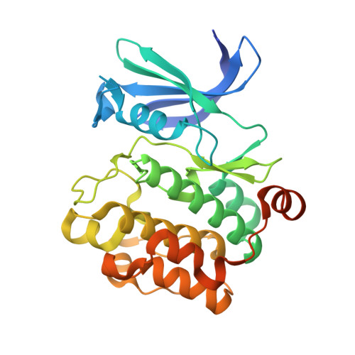
    3UMW

  • PubMed Abstract: 

    Serine/threonine kinase PIM1 is an emerging therapeutic target for hematopoietic and prostate cancer therapy. To develop a novel PIM1 inhibitor, we focused on 1, a metabolically labile, nonselective kinase inhibitor discovered in our previous screening study. We adopted a rational optimization strategy based mainly on structural information for the PIM1-1 complex to improve the potency and selectivity. This approach afforded the potent and metabolically stable PIM1-selective inhibitor 14, which shows only a marginal increase in molecular weight compared with 1 but has a significantly decreased cLogP. The validity of our design concept was confirmed by X-ray structure analysis. In a cellular study, 14 potently inhibited the growth of human leukemia cell line MV4-11 but had a negligible effect on the growth of WI-38 (surrogate for general toxicity). These results demonstrate the effectiveness of our design strategy for evolving the screening-hit compound 1 into a novel type of PIM1 inhibitor, 14.


  • Organizational Affiliation
    • Open Innovation Center for Drug Discovery, The University of Tokyo, 7-3-1 Hongo, Bunkyo-ku, Tokyo 113-0033, Japan.

Macromolecule Content 

  • Total Structure Weight: 34.77 kDa 
  • Atom Count: 2,429 
  • Modeled Residue Count: 264 
  • Deposited Residue Count: 298 
  • Unique protein chains: 1

Macromolecules

Find similar proteins by:|  3D Structure
Entity ID: 1
MoleculeChains  Sequence LengthOrganismDetailsImage
Proto-oncogene serine/threonine-protein kinase pim-1298Homo sapiensMutation(s): 0 
Gene Names: pim1
EC: 2.7.11.1
UniProt & NIH Common Fund Data Resources
Find proteins for P11309 (Homo sapiens)
Explore P11309 
Go to UniProtKB:  P11309
PHAROS:  P11309
GTEx:  ENSG00000137193 
Entity Groups
Sequence Clusters30% Identity50% Identity70% Identity90% Identity95% Identity100% Identity
UniProt GroupP11309
Sequence Annotations
Expand
Reference Sequence

Small Molecules

Binding Affinity Annotations 
IDSourceBinding Affinity
596 BindingDB:  3UMW IC50: min: 3, max: 6 (nM) from 2 assay(s)

Experimental Data & Validation

Experimental Data

  • Method: X-RAY DIFFRACTION
  • Resolution: 2.08 Å
  • R-Value Free:  0.200 (Depositor), 0.200 (DCC) 
  • R-Value Work:  0.173 (Depositor), 0.174 (DCC) 
  • R-Value Observed: 0.175 (Depositor) 
Space Group: P 65
Unit Cell:
Length ( Å )Angle ( ˚ )
a = 98.28α = 90
b = 98.28β = 90
c = 80.641γ = 120
Software Package:
Software NamePurpose
SCALAdata scaling
PHASERphasing
REFMACrefinement
PDB_EXTRACTdata extraction
HKL-2000data collection
MOSFLMdata reduction

Structure Validation

View Full Validation Report


Ligand Structure Quality Assessment 


Entry History 

Deposition Data

Revision History  (Full details and data files)

  • Version 1.0: 2012-10-03
    Type: Initial release
  • Version 1.1: 2023-11-01
    Changes: Data collection, Database references, Derived calculations, Refinement description