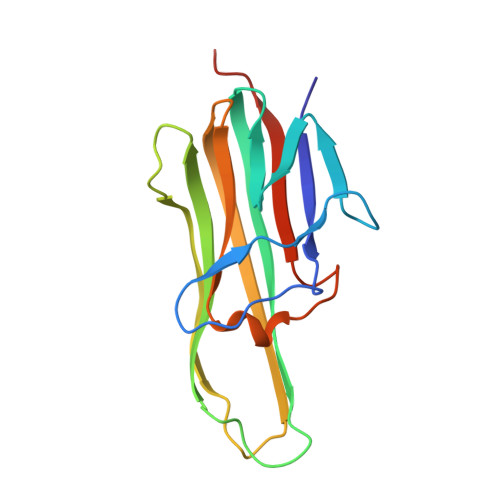
3URF

Human RANKL/OPG complex


Experimental Data Snapshot

  • Method: X-RAY DIFFRACTION
  • Resolution: 2.70 Å
  • R-Value Free: 0.251 
  • R-Value Work: 0.202 
  • R-Value Observed: 0.205 

wwPDB Validation   3D Report Full Report


Ligand Structure Quality Assessment 


This is version 1.2 of the entry. See complete history


Literature

Crystal Structure of Human RANKL Complexed with Its Decoy Receptor Osteoprotegerin

Luan, X.D.Lu, Q.Y.Jiang, Y.N.Zhang, S.Y.Wang, Q.Yuan, H.H.Zhao, W.M.Wang, J.W.Wang, X.Q.

(2012) J Immunol 189: 245-252

  • DOI: https://doi.org/10.4049/jimmunol.1103387
  • Primary Citation of Related Structures:  
    3URF

  • PubMed Abstract: 

    Receptor activator of NF-κB ligand (RANKL), its signaling receptor RANK, and its decoy receptor osteoprotegerin (OPG) constitute a molecular triad that is critical in regulating bone remodeling, and also plays multiple roles in the immune system. OPG binds RANKL directly to block its interaction with RANK. In this article, we report the 2.7-Å crystal structure of human RANKL trimer in complex with the N-terminal fragment of human OPG containing four cysteine-rich TNFR homologous domains (OPG-CRD). The structure shows that RANKL trimer uses three equivalent grooves between two neighboring monomers to interact with three OPG-CRD monomers symmetrically. A loop from the CRD3 domain of OPG-CRD inserts into the shallow groove of RANKL, providing the major binding determinant that is further confirmed by affinity measurement and osteoclast differentiation assay. These results, together with a previously reported mouse RANKL/RANK complex structure, reveal that OPG exerts its decoy receptor function by directly blocking the accessibilities of important interacting residues of RANKL for RANK recognition. Structural comparison with TRAIL/death receptor 5 complex also reveals structural basis for the cross-reactivity of OPG to TRAIL.


  • Organizational Affiliation

    Center for Structural Biology, School of Life Sciences, Ministry of Education Key Laboratory of Protein Science, Tsinghua University, Beijing 100084, People's Republic of China.


Macromolecules
Find similar proteins by:  (by identity cutoff)  |  3D Structure
Entity ID: 1
MoleculeChains Sequence LengthOrganismDetailsImage
Tumor necrosis factor ligand superfamily member 11, soluble form162Homo sapiensMutation(s): 0 
Gene Names: TNFSF11OPGLRANKLTRANCE
UniProt & NIH Common Fund Data Resources
Find proteins for O14788 (Homo sapiens)
Explore O14788 
Go to UniProtKB:  O14788
PHAROS:  O14788
GTEx:  ENSG00000120659 
Entity Groups  
Sequence Clusters30% Identity50% Identity70% Identity90% Identity95% Identity100% Identity
UniProt GroupO14788
Sequence Annotations
Expand
  • Reference Sequence
Find similar proteins by:  (by identity cutoff)  |  3D Structure
Entity ID: 2
MoleculeChains Sequence LengthOrganismDetailsImage
Tumor necrosis factor receptor superfamily member 11BB [auth Z]171Homo sapiensMutation(s): 0 
Gene Names: TNFRSF11BOCIFOPG
UniProt & NIH Common Fund Data Resources
Find proteins for O00300 (Homo sapiens)
Explore O00300 
Go to UniProtKB:  O00300
PHAROS:  O00300
GTEx:  ENSG00000164761 
Entity Groups  
Sequence Clusters30% Identity50% Identity70% Identity90% Identity95% Identity100% Identity
UniProt GroupO00300
Glycosylation
Glycosylation Sites: 1Go to GlyGen: O00300-1
Sequence Annotations
Expand
  • Reference Sequence
Small Molecules
Ligands 1 Unique
IDChains Name / Formula / InChI Key2D Diagram3D Interactions
NAG
Query on NAG

Download Ideal Coordinates CCD File 
C [auth Z]2-acetamido-2-deoxy-beta-D-glucopyranose
C8 H15 N O6
OVRNDRQMDRJTHS-FMDGEEDCSA-N
Experimental Data & Validation

Experimental Data

  • Method: X-RAY DIFFRACTION
  • Resolution: 2.70 Å
  • R-Value Free: 0.251 
  • R-Value Work: 0.202 
  • R-Value Observed: 0.205 
  • Space Group: P 41 3 2
Unit Cell:
Length ( Å )Angle ( ˚ )
a = 144.8α = 90
b = 144.8β = 90
c = 144.8γ = 90
Software Package:
Software NamePurpose
ADSCdata collection
PHASERphasing
PHENIXrefinement
MOSFLMdata reduction
SCALAdata scaling

Structure Validation

View Full Validation Report


Ligand Structure Quality Assessment 


Entry History 

Deposition Data

Revision History  (Full details and data files)

  • Version 1.0: 2012-07-11
    Type: Initial release
  • Version 1.1: 2020-07-29
    Type: Remediation
    Reason: Carbohydrate remediation
    Changes: Data collection, Database references, Derived calculations, Structure summary
  • Version 1.2: 2024-10-30
    Changes: Data collection, Database references, Structure summary