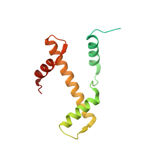
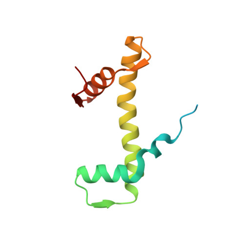
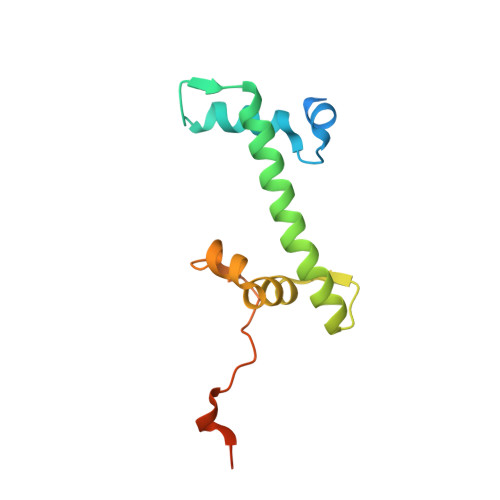
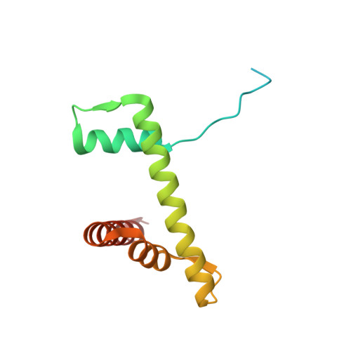
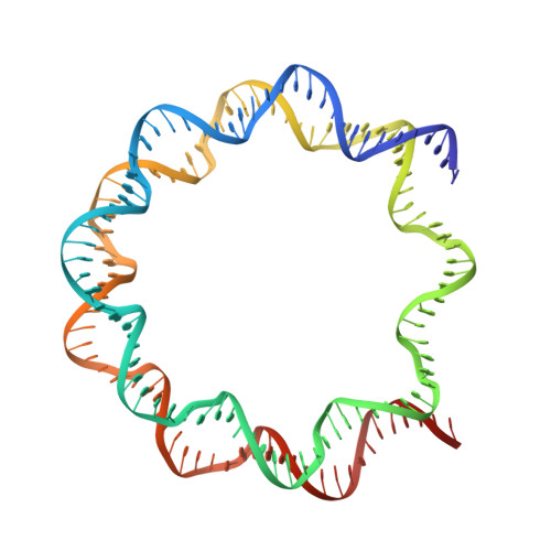
3UTB

Crystal Structure of Nucleosome Core Particle Assembled with the 146b Alpha-Satellite Sequence (NCP146b)


Experimental Data Snapshot

  • Method: X-RAY DIFFRACTION
  • Resolution: 2.20 Å
  • R-Value Free: 0.275 
  • R-Value Work: 0.232 
  • R-Value Observed: 0.233 

wwPDB Validation   3D Report Full Report


This is version 1.2 of the entry. See complete history


Literature

The mechanics behind DNA sequence-dependent properties of the nucleosome

Chua, E.Y.D.Vasudevan, D.Davey, G.E.Wu, B.Davey, C.A.

(2012) Nucleic Acids Res 40: 6338-6352

  • DOI: https://doi.org/10.1093/nar/gks261
  • Primary Citation of Related Structures:  
    3UT9, 3UTA, 3UTB

  • PubMed Abstract: 

    Chromatin organization and composition impart sophisticated regulatory features critical to eukaryotic genomic function. Although DNA sequence-dependent histone octamer binding is important for nucleosome activity, many aspects of this phenomenon have remained elusive. We studied nucleosome structure and stability with diverse DNA sequences, including Widom 601 derivatives with the highest known octamer affinities, to establish a simple model behind the mechanics of sequence dependency. This uncovers the unique but unexpected role of TA dinucleotides and a propensity for G|C-rich sequence elements to conform energetically favourably at most locations around the histone octamer, which rationalizes G|C% as the most predictive factor for nucleosome occupancy in vivo. In addition, our findings reveal dominant constraints on double helix conformation by H3-H4 relative to H2A-H2B binding and DNA sequence context-dependency underlying nucleosome structure, positioning and stability. This provides a basis for improved prediction of nucleosomal properties and the design of tailored DNA constructs for chromatin investigations.


  • Organizational Affiliation

    Division of Structural and Computational Biology, School of Biological Sciences, Nanyang Technological University, 60 Nanyang Drive, Singapore 637551, Singapore.


Macromolecules

Find similar proteins by:  (by identity cutoff)  |  3D Structure
Entity ID: 1
MoleculeChains Sequence LengthOrganismDetailsImage
Histone H3.2
A, E
135Xenopus laevisMutation(s): 0 
UniProt
Find proteins for P84233 (Xenopus laevis)
Explore P84233 
Go to UniProtKB:  P84233
Entity Groups  
Sequence Clusters30% Identity50% Identity70% Identity90% Identity95% Identity100% Identity
UniProt GroupP84233
Sequence Annotations
Expand
  • Reference Sequence
Find similar proteins by:  (by identity cutoff)  |  3D Structure
Entity ID: 2
MoleculeChains Sequence LengthOrganismDetailsImage
Histone H4
B, F
102Xenopus laevisMutation(s): 0 
UniProt
Find proteins for P62799 (Xenopus laevis)
Explore P62799 
Go to UniProtKB:  P62799
Entity Groups  
Sequence Clusters30% Identity50% Identity70% Identity90% Identity95% Identity100% Identity
UniProt GroupP62799
Sequence Annotations
Expand
  • Reference Sequence
Find similar proteins by:  (by identity cutoff)  |  3D Structure
Entity ID: 3
MoleculeChains Sequence LengthOrganismDetailsImage
Histone H2A
C, G
129Xenopus laevisMutation(s): 0 
Gene Names: hist1h2ajLOC494591
UniProt
Find proteins for P06897 (Xenopus laevis)
Explore P06897 
Go to UniProtKB:  P06897
Entity Groups  
Sequence Clusters30% Identity50% Identity70% Identity90% Identity95% Identity100% Identity
UniProt GroupP06897
Sequence Annotations
Expand
  • Reference Sequence
Find similar proteins by:  (by identity cutoff)  |  3D Structure
Entity ID: 4
MoleculeChains Sequence LengthOrganismDetailsImage
Histone H2B 1.1
D, H
125Xenopus laevisMutation(s): 0 
UniProt
Find proteins for P02281 (Xenopus laevis)
Explore P02281 
Go to UniProtKB:  P02281
Entity Groups  
Sequence Clusters30% Identity50% Identity70% Identity90% Identity95% Identity100% Identity
UniProt GroupP02281
Sequence Annotations
Expand
  • Reference Sequence
Find similar nucleic acids by:  (by identity cutoff)  |  3D Structure
Entity ID: 5
MoleculeChains LengthOrganismImage
146-mer DNA
I, J
146N/A
Sequence Annotations
Expand
  • Reference Sequence
Small Molecules
Ligands 2 Unique
IDChains Name / Formula / InChI Key2D Diagram3D Interactions
SO4
Query on SO4

Download Ideal Coordinates CCD File 
L [auth C],
N [auth D],
P [auth G]
SULFATE ION
O4 S
QAOWNCQODCNURD-UHFFFAOYSA-L
MN
Query on MN

Download Ideal Coordinates CCD File 
AA [auth J]
BA [auth J]
CA [auth J]
DA [auth J]
EA [auth J]
AA [auth J],
BA [auth J],
CA [auth J],
DA [auth J],
EA [auth J],
FA [auth J],
GA [auth J],
HA [auth J],
K [auth A],
M [auth D],
O [auth E],
Q [auth I],
R [auth I],
S [auth I],
T [auth I],
U [auth I],
V [auth I],
W [auth I],
X [auth I],
Y [auth I],
Z [auth J]
MANGANESE (II) ION
Mn
WAEMQWOKJMHJLA-UHFFFAOYSA-N
Experimental Data & Validation

Experimental Data

  • Method: X-RAY DIFFRACTION
  • Resolution: 2.20 Å
  • R-Value Free: 0.275 
  • R-Value Work: 0.232 
  • R-Value Observed: 0.233 
  • Space Group: P 21 21 21
Unit Cell:
Length ( Å )Angle ( ˚ )
a = 105.46α = 90
b = 109.28β = 90
c = 175.92γ = 90
Software Package:
Software NamePurpose
SCALAdata scaling
REFMACrefinement
PDB_EXTRACTdata extraction

Structure Validation

View Full Validation Report


Entry History 

Deposition Data

Revision History  (Full details and data files)

  • Version 1.0: 2012-04-11
    Type: Initial release
  • Version 1.1: 2013-06-26
    Changes: Database references
  • Version 1.2: 2024-03-20
    Changes: Data collection, Database references, Derived calculations