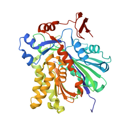
3V4N

The Biochemical and Structural Basis for Inhibition of Enterococcus faecalis HMG-CoA Synthatse, mvaS, by Hymeglusin


Experimental Data Snapshot

  • Method: X-RAY DIFFRACTION
  • Resolution: 1.60 Å
  • R-Value Free: 0.194 
  • R-Value Work: 0.141 
  • R-Value Observed: 0.146 

wwPDB Validation   3D Report Full Report


This is version 1.3 of the entry. See complete history


Literature

Biochemical and structural basis for inhibition of Enterococcus faecalis hydroxymethylglutaryl-CoA synthase, mvaS, by hymeglusin.

Skaff, D.A.Ramyar, K.X.McWhorter, W.J.Barta, M.L.Geisbrecht, B.V.Miziorko, H.M.

(2012) Biochemistry 51: 4713-4722

  • DOI: https://doi.org/10.1021/bi300037k
  • Primary Citation of Related Structures:  
    3V4N, 3V4X

  • PubMed Abstract: 

    Hymeglusin (1233A, F244, L-659-699) is established as a specific β-lactone inhibitor of eukaryotic hydroxymethylglutaryl-CoA synthase (HMGCS). Inhibition results from formation of a thioester adduct to the active site cysteine. In contrast, the effects of hymeglusin on bacterial HMG-CoA synthase, mvaS, have been minimally characterized. Hymeglusin blocks growth of Enterococcus faecalis. After removal of the inhibitor from culture media, a growth curve inflection point at 3.1 h is observed (vs 0.7 h for the uninhibited control). Upon hymeglusin inactivation of purified E. faecalis mvaS, the thioester adduct is more stable than that measured for human HMGCS. Hydroxylamine cleaves the thioester adduct; substantial enzyme activity is restored at a rate that is 8-fold faster for human HMGCS than for mvaS. Structural results explain these differences in enzyme-inhibitor thioester adduct stability and solvent accessibility. The E. faecalis mvaS-hymeglusin cocrystal structure (1.95 Å) reveals virtually complete occlusion of the bound inhibitor in a narrow tunnel that is largely sequestered from bulk solvent. In contrast, eukaryotic (Brassica juncea) HMGCS binds hymeglusin in a more solvent-exposed cavity.


  • Organizational Affiliation

    University of Missouri, Kansas City, MO, USA.


Macromolecules
Find similar proteins by:  (by identity cutoff)  |  3D Structure
Entity ID: 1
MoleculeChains Sequence LengthOrganismDetailsImage
HMG-CoA synthase
A, B, C, D
388Enterococcus faecalisMutation(s): 1 
Gene Names: mvaS
EC: 2.3.3.10
UniProt
Find proteins for Q9FD71 (Enterococcus faecalis)
Explore Q9FD71 
Go to UniProtKB:  Q9FD71
Entity Groups  
Sequence Clusters30% Identity50% Identity70% Identity90% Identity95% Identity100% Identity
UniProt GroupQ9FD71
Sequence Annotations
Expand
  • Reference Sequence
Small Molecules
Ligands 1 Unique
IDChains Name / Formula / InChI Key2D Diagram3D Interactions
BTB
Query on BTB

Download Ideal Coordinates CCD File 
E [auth B]2-[BIS-(2-HYDROXY-ETHYL)-AMINO]-2-HYDROXYMETHYL-PROPANE-1,3-DIOL
C8 H19 N O5
OWMVSZAMULFTJU-UHFFFAOYSA-N
Modified Residues  1 Unique
IDChains TypeFormula2D DiagramParent
SNC
Query on SNC
A, B, C, D
L-PEPTIDE LINKINGC3 H6 N2 O3 SCYS
Experimental Data & Validation

Experimental Data

  • Method: X-RAY DIFFRACTION
  • Resolution: 1.60 Å
  • R-Value Free: 0.194 
  • R-Value Work: 0.141 
  • R-Value Observed: 0.146 
  • Space Group: P 1 21 1
Unit Cell:
Length ( Å )Angle ( ˚ )
a = 109.531α = 90
b = 56.279β = 100.02
c = 123.893γ = 90
Software Package:
Software NamePurpose
HKL-2000data collection
PHENIXmodel building
PHENIXrefinement
HKL-2000data reduction
HKL-2000data scaling
PHENIXphasing

Structure Validation

View Full Validation Report


Entry History 

Deposition Data

Revision History  (Full details and data files)

  • Version 1.0: 2012-04-25
    Type: Initial release
  • Version 1.1: 2013-01-30
    Changes: Database references
  • Version 1.2: 2014-09-10
    Changes: Refinement description
  • Version 1.3: 2024-10-16
    Changes: Data collection, Database references, Derived calculations, Structure summary