Design and evaluation of azaindole-substituted N-hydroxypyridones as glyoxalase I inhibitors
Chiba, T., Ohwada, J., Sakamoto, H., Kobayashi, T., Fukami, T.A., Irie, M., Miura, T., Ohara, K., Koyano, H.(2012) Bioorg Med Chem Lett 22: 7486-7489
- PubMed: 23122816 Search on PubMed
- DOI: https://doi.org/10.1016/j.bmcl.2012.10.045
- Primary Citation Related Structures:
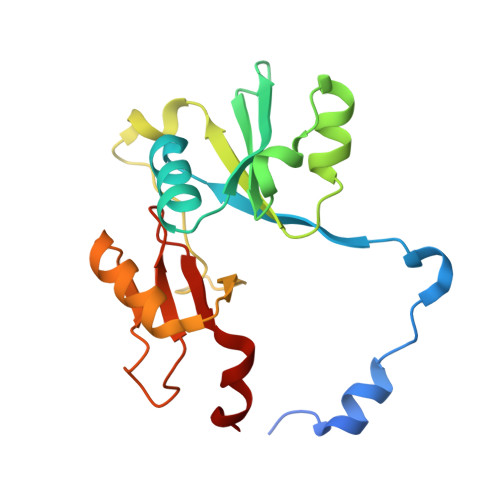
3VW9 - PubMed Abstract:
We conducted a high throughput screening for glyoxalase I (GLO1) inhibitors and identified 4,6-diphenyl-N-hydroxypyridone as a lead compound. Using a binding model of the lead and public X-ray coordinates of GLO1 enzymes complexed with glutathione analogues, we designed 4-(7-azaindole)-substituted 6-phenyl-N-hydroxypyridones. 7-Azaindole's 7-nitrogen was expected to interact with a water network, resulting in an interaction with the protein. We validated this inhibitor design by comparing its structure-activity relationship (SAR) with that of corresponding indole derivatives, by analyzing the binding mode with X-ray crystallography and by evaluating its thermodynamic binding parameters.
- Research Division, Chugai Pharmaceutical Co., Ltd, 200 Kajiwara Kamakura, Kanagawa 247-8530, Japan.
Organizational Affiliation: