Lead optimization of novel p53-MDM2 interaction inhibitors possessing dihydroimidazothiazole scaffold
Miyazaki, M., Naito, H., Sugimoto, Y., Kawato, H., Okayama, T., Shimizu, H., Miyazaki, M., Kitagawa, M., Seki, T., Fukutake, S., Aonuma, M., Soga, T.(2013) Bioorg Med Chem Lett 23: 728-732
- PubMed: 23266121 Search on PubMed
- DOI: https://doi.org/10.1016/j.bmcl.2012.11.091
- Primary Citation Related Structures:
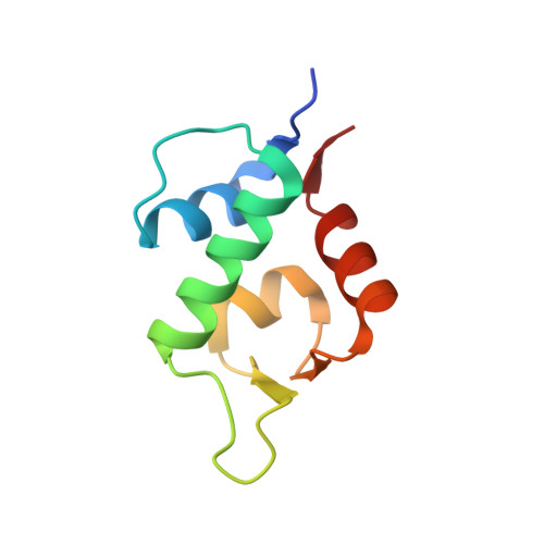
3VZV - PubMed Abstract:
With the aim of discovering potent inhibitors of the p53-MDM2 interaction and thus obtaining a potent anticancer drug, we have pursued synthesis and optimization of dihydroimidazothiazole derivatives, which have been discovered via scaffold hopping by mimicing the mode of interaction between MDM2 and Nutlins. Upon the discovery we encountered a problem involving the chemical instability of the scaffold, that is, susceptibility to oxidation which led to imidazothiazole. In order to solve this problem and to obtain further potent compounds, we executed medicinal research and thus furnished the optimal compounds by incorporating the methyl group onto the C-6 position to avoid the oxidation, and by modifying the C-2 moiety of the additional proline motif, which furnished high potency. The incorporation of the pyrrolidine moiety at the C-2 position raised another hydrophobic interaction site with MDM2 protein, which was generated by the induced-fitting observed by co-crystal structure analysis. These optimal molecules showed significant improvement in potency when compared with the early lead (+)-1 or Nutlin-3a.
- Lead Discovery & Optimization Research Laboratories II, R&D Division, Daiichi Sankyo Co., Ltd, 1-2-58 Hiromachi, Tokyo 140-8710, Japan. miyazaki.masaki.p3@daiichisankyo.co.jp
Organizational Affiliation: