3W5E | pdb_00003w5e

Crystal structure of phosphodiesterase 4B in complex with compound 31e


Experimental Data Snapshot

  • Method: X-RAY DIFFRACTION
  • Resolution: 2.30 Å
  • R-Value Free: 
    0.269 (Depositor) 
  • R-Value Work: 
    0.182 (Depositor) 
  • R-Value Observed: 
    0.190 (Depositor) 

Starting Model: experimental
View more details

wwPDB Validation 3D Report Full Report

Validation slider image for 3W5E

Ligand Structure Quality Assessment 


This is version 1.3 of the entry. See complete history

Literature

Identification of the fused bicyclic 4-amino-2-phenylpyrimidine derivatives as novel and potent PDE4 inhibitors

Goto, T.Shiina, A.Yoshino, T.Mizukami, K.Hirahara, K.Suzuki, O.Sogawa, Y.Takahashi, T.Mikkaichi, T.Nakao, N.Takahashi, M.Hasegawa, M.Sasaki, S.

(2013) Bioorg Med Chem Lett 23: 3325-3328

  • DOI: https://doi.org/10.1016/j.bmcl.2013.03.104
  • Primary Citation Related Structures: 
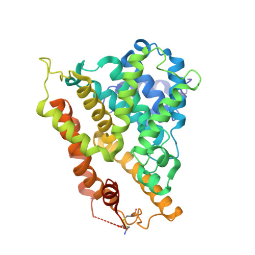
    3W5E

  • PubMed Abstract: 

    2-Phenyl-4-piperidinyl-6,7-dihydrothieno[3,4-d]pyrimidine derivative (2) was found to be a new PDE4 inhibitor with moderate PDE4B activity (IC50=150 nM). A number of derivatives with a variety of 4-amino substituents and fused bicyclic pyrimidines were synthesized. Among these, 5,5-dioxo-7,8-dihydro-6H-thiopyrano[3,2-d]pyrimidine derivative (18) showed potent PDE4B inhibitory activity (IC50=25 nM). Finally, N-propylacetamide derivative (31b) was determined as a potent inhibitor for both PDE4B (IC50=7.5 nM) and TNF-α production in mouse splenocytes (IC50=9.8 nM) and showed good in vivo anti-inflammatory activity in the LPS-induced lung inflammation model in mice (ID50=18 mg/kg). The binding mode of the new inhibitor (31e) in the catalytic site of PDE4B is presented based on an X-ray crystal structure of the ligand-enzyme complex.


  • Organizational Affiliation
    • R&D Division, Daiichi Sankyo Co., Ltd, 1-2-58 Hiromachi, Tokyo 140-8710, Japan. goto.taiji.kt@daiichisankyo.co.jp

Macromolecule Content 

  • Total Structure Weight: 87.85 kDa 
  • Atom Count: 5,994 
  • Modeled Residue Count: 690 
  • Deposited Residue Count: 754 
  • Unique protein chains: 1

Macromolecules

Find similar proteins by:|  3D Structure
Entity ID: 1
MoleculeChains  Sequence LengthOrganismDetailsImage
cAMP-specific 3',5'-cyclic phosphodiesterase 4B
A, B
377Homo sapiensMutation(s): 0 
Gene Names: DPDE4PDE4B
EC: 3.1.4.17 (PDB Primary Data), 3.1.4.53 (UniProt)
UniProt & NIH Common Fund Data Resources
Find proteins for Q07343 (Homo sapiens)
Explore Q07343 
Go to UniProtKB:  Q07343
PHAROS:  Q07343
GTEx:  ENSG00000184588 
Entity Groups
Sequence Clusters30% Identity50% Identity70% Identity90% Identity95% Identity100% Identity
UniProt GroupQ07343
Sequence Annotations
Expand
Reference Sequence

Small Molecules

Ligands 3 Unique
IDChains Name / Formula / InChI Key2D Diagram3D Interactions
NVW

Query on NVW


Download:Ideal Coordinates CCD File
C [auth A],
F [auth B]
N-tert-butyl-2-{4-[(5,5-dioxido-2-phenyl-7,8-dihydro-6H-thiopyrano[3,2-d]pyrimidin-4-yl)amino]phenyl}acetamide
C25 H28 N4 O3 S
IHIZFBWFXODXJS-UHFFFAOYSA-N
ZN

Query on ZN


Download:Ideal Coordinates CCD File
D [auth A],
G [auth B]
ZINC ION
Zn
PTFCDOFLOPIGGS-UHFFFAOYSA-N
CA

Query on CA


Download:Ideal Coordinates CCD File
E [auth A],
H [auth B]
CALCIUM ION
Ca
BHPQYMZQTOCNFJ-UHFFFAOYSA-N

Experimental Data & Validation

Experimental Data

  • Method: X-RAY DIFFRACTION
  • Resolution: 2.30 Å
  • R-Value Free:  0.269 (Depositor) 
  • R-Value Work:  0.182 (Depositor) 
  • R-Value Observed: 0.190 (Depositor) 
Space Group: P 21 21 2
Unit Cell:
Length ( Å )Angle ( ˚ )
a = 81.064α = 90
b = 158.19β = 90
c = 58.157γ = 90
Software Package:
Software NamePurpose
DENZOdata reduction
SCALEPACKdata scaling
REFMACrefinement
PDB_EXTRACTdata extraction
HKL-2000data reduction
HKL-2000data scaling
CNSphasing

Structure Validation

View Full Validation Report


Ligand Structure Quality Assessment 


Entry History 

Deposition Data

Revision History  (Full details and data files)

  • Version 1.0: 2013-05-29
    Type: Initial release
  • Version 1.1: 2017-11-22
    Changes: Refinement description
  • Version 1.2: 2023-11-08
    Changes: Data collection, Database references, Derived calculations, Refinement description
  • Version 1.3: 2024-11-13
    Changes: Structure summary