Conformational restriction approach to beta-secretase (BACE1) inhibitors III: Effective investigation of the binding mode by combinational use of X-ray analysis, isothermal titration calorimetry and theoretical calculations
Yonezawa, S., Fujiwara, K., Yamamoto, T., Hattori, K., Yamakawa, H., Muto, C., Hosono, M., Tanaka, Y., Nakano, T., Takemoto, H., Arisawa, M., Shuto, S.(2013) Bioorg Med Chem 21: 6506-6522
- PubMed: 24051074 Search on PubMed
- DOI: https://doi.org/10.1016/j.bmc.2013.08.036
- Primary Citation Related Structures:
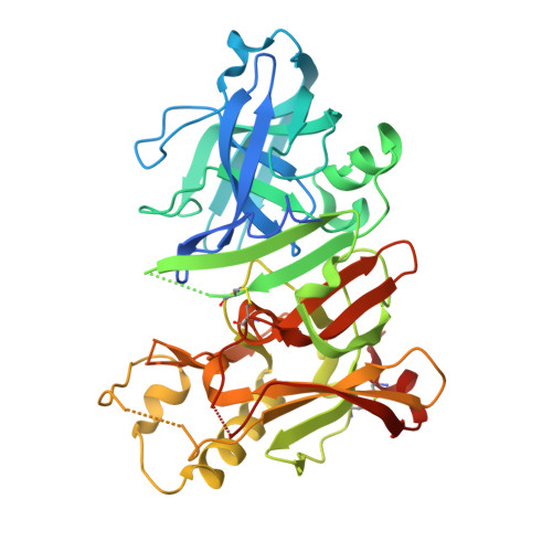
3WB4, 3WB5 - PubMed Abstract:
For further investigation of BACE1 inhibitors using conformational restriction with sp(3) hybridized carbon, we applied this approach to 6-substituted aminopyrimidone derivatives 3 to improve the inhibitory activity by reducing the entropic energy loss upon binding to BACE1. Among eight stereoisomers synthesized, [trans-(1'R,2'R),6S] isomer 6 exhibited the best BACE1 inhibitory activity, which was statistically superior to that of the corresponding ethylene linker compound (R)-3. Combinational examinations of the binding mode of 6 were performed, which included isothermal titration calorimetry (ITC), X-ray crystallographic structure analysis and theoretical calculations, to clarify the effect of our conformational restriction approach. From the ITC measurement, the binding entropy of 6 was found to be ∼0.5kcal larger than that of (R)-3, which is considered to be affected by conformational restriction with a cyclopropane ring.
- Shionogi Innovation Center for Drug Discovery, Shionogi & Co., Ltd, Kita-21 Nishi-11 Kita-ku, Sapporo 001-0021, Japan; Faculty of Pharmaceutical Sciences, Hokkaido University, Kita-12, Nishi-6, Kita-ku, Sapporo 060-0812, Japan. Electronic address: shuji.yonezawa@shionogi.co.jp.
Organizational Affiliation: