


Crystal structure of GYE (old yellow enzyme)
Yuan, Y.A., Yin, B.To be published.
Experimental Data Snapshot
wwPDB Validation 3D Report Full Report

Entity ID: 1 | |||||
|---|---|---|---|---|---|
| Molecule | Chains | Sequence Length | Organism | Details | Image |
| NADH oxidase | 361 | Gluconobacter oxydans | Mutation(s): 0 Gene Names: nox | 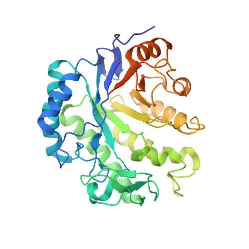 | |
UniProt | |||||
Entity Groups | |||||
| Sequence Clusters | 30% Identity50% Identity70% Identity90% Identity95% Identity100% Identity | ||||
| UniProt Group | A1E8I9 | ||||
Sequence AnnotationsExpand | |||||
Reference Sequence | |||||
| Ligands 3 Unique | |||||
|---|---|---|---|---|---|
| ID | Chains | Name / Formula / InChI Key | 2D Diagram | 3D Interactions | |
| HG Download:Ideal Coordinates CCD File | D [auth A] | MERCURY (II) ION Hg BQPIGGFYSBELGY-UHFFFAOYSA-N |  | ||
| PGE Download:Ideal Coordinates CCD File | B [auth A] | TRIETHYLENE GLYCOL C6 H14 O4 ZIBGPFATKBEMQZ-UHFFFAOYSA-N |  | ||
| PEG Download:Ideal Coordinates CCD File | C [auth A] | DI(HYDROXYETHYL)ETHER C4 H10 O3 MTHSVFCYNBDYFN-UHFFFAOYSA-N |  | ||
| Length ( Å ) | Angle ( ˚ ) |
|---|---|
| a = 138.886 | α = 90 |
| b = 138.886 | β = 90 |
| c = 145.091 | γ = 120 |
| Software Name | Purpose |
|---|---|
| HKL-2000 | data collection |
| SHARP | phasing |
| REFMAC | refinement |
| HKL-2000 | data reduction |
| HKL-2000 | data scaling |


