3WO3

Crystal structure of IL-18 in complex with IL-18 receptor alpha


Experimental Data Snapshot

  • Method: X-RAY DIFFRACTION
  • Resolution: 3.10 Å
  • R-Value Free: 0.222 
  • R-Value Work: 0.191 
  • R-Value Observed: 0.192 

wwPDB Validation   3D Report Full Report


Ligand Structure Quality Assessment 


This is version 2.1 of the entry. See complete history


Literature

The structural basis for receptor recognition of human interleukin-18

Tsutsumi, N.Kimura, T.Arita, K.Ariyoshi, M.Ohnishi, H.Yamamoto, T.Zuo, X.Maenaka, K.Park, E.Y.Kondo, N.Shirakawa, M.Tochio, H.Kato, Z.

(2014) Nat Commun 5: 5340-5340

  • DOI: https://doi.org/10.1038/ncomms6340
  • Primary Citation of Related Structures:  
    3WO2, 3WO3, 3WO4

  • PubMed Abstract: 

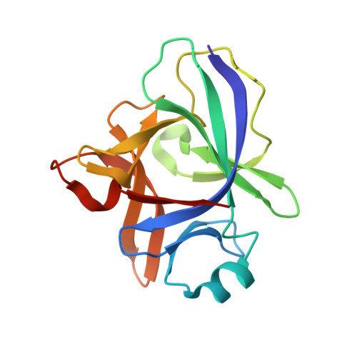
    Interleukin (IL)-18 is a proinflammatory cytokine that belongs to the IL-1 family and plays an important role in inflammation. The uncontrolled release of this cytokine is associated with severe chronic inflammatory disease. IL-18 forms a signalling complex with the IL-18 receptor α (Rα) and β (Rβ) chains at the plasma membrane, which induces multiple inflammatory cytokines. Here, we present a crystal structure of human IL-18 bound to the two receptor extracellular domains. Generally, the receptors' recognition mode for IL-18 is similar to IL-1β; however, certain notable differences were observed. The architecture of the IL-18 receptor second domain (D2) is unique among the other IL-1R family members, which presumably distinguishes them from the IL-1 receptors that exhibit a more promiscuous ligand recognition mode. The structures and associated biochemical and cellular data should aid in developing novel drugs to neutralize IL-18 activity.


  • Organizational Affiliation

    Department of Molecular Engineering, Graduate School of Engineering, Kyoto University, Katsura, Nishikyo-ku, Kyoto 615-8510, Japan.


Macromolecules
Find similar proteins by:  (by identity cutoff)  |  3D Structure
Entity ID: 1
MoleculeChains Sequence LengthOrganismDetailsImage
Interleukin-18
A, C, E, G, I
A, C, E, G, I, K
157Homo sapiensMutation(s): 0 
Gene Names: IL18
UniProt & NIH Common Fund Data Resources
Find proteins for Q14116 (Homo sapiens)
Explore Q14116 
Go to UniProtKB:  Q14116
PHAROS:  Q14116
GTEx:  ENSG00000150782 
Entity Groups  
Sequence Clusters30% Identity50% Identity70% Identity90% Identity95% Identity100% Identity
UniProt GroupQ14116
Sequence Annotations
Expand
  • Reference Sequence
Find similar proteins by:  (by identity cutoff)  |  3D Structure
Entity ID: 2
MoleculeChains Sequence LengthOrganismDetailsImage
Interleukin-18 receptor 1
B, D, F, H, J
B, D, F, H, J, L
312Homo sapiensMutation(s): 0 
Gene Names: IL18R1
UniProt & NIH Common Fund Data Resources
Find proteins for Q13478 (Homo sapiens)
Explore Q13478 
Go to UniProtKB:  Q13478
PHAROS:  Q13478
GTEx:  ENSG00000115604 
Entity Groups  
Sequence Clusters30% Identity50% Identity70% Identity90% Identity95% Identity100% Identity
UniProt GroupQ13478
Sequence Annotations
Expand
  • Reference Sequence
Oligosaccharides

Help

Entity ID: 3
MoleculeChains Length2D Diagram Glycosylation3D Interactions
2-acetamido-2-deoxy-beta-D-glucopyranose-(1-4)-[alpha-L-fucopyranose-(1-6)]2-acetamido-2-deoxy-beta-D-glucopyranoseHA [auth h],
M,
Q
3N-Glycosylation
Glycosylation Resources
GlyTouCan:  G21290RB
GlyCosmos:  G21290RB
GlyGen:  G21290RB
Entity ID: 4
MoleculeChains Length2D Diagram Glycosylation3D Interactions
alpha-D-mannopyranose-(1-6)-alpha-D-mannopyranose-(1-6)-[alpha-D-mannopyranose-(1-3)]beta-D-mannopyranose-(1-4)-2-acetamido-2-deoxy-beta-D-glucopyranose-(1-4)-2-acetamido-2-deoxy-beta-D-glucopyranose
N, W
6N-Glycosylation
Glycosylation Resources
GlyTouCan:  G34442SS
GlyCosmos:  G34442SS
GlyGen:  G34442SS
Entity ID: 5
MoleculeChains Length2D Diagram Glycosylation3D Interactions
2-acetamido-2-deoxy-beta-D-glucopyranose-(1-4)-2-acetamido-2-deoxy-beta-D-glucopyranoseBA [auth b],
GA [auth g],
JA [auth j],
KA [auth k],
O
2N-Glycosylation
Glycosylation Resources
GlyTouCan:  G42666HT
GlyCosmos:  G42666HT
GlyGen:  G42666HT
Entity ID: 6
MoleculeChains Length2D Diagram Glycosylation3D Interactions
beta-D-mannopyranose-(1-4)-2-acetamido-2-deoxy-beta-D-glucopyranose-(1-4)-2-acetamido-2-deoxy-beta-D-glucopyranose
CA [auth c],
EA [auth e],
FA [auth f],
IA [auth i],
P,
CA [auth c],
EA [auth e],
FA [auth f],
IA [auth i],
P,
T,
U,
X
3N-Glycosylation
Glycosylation Resources
GlyTouCan:  G15407YE
GlyCosmos:  G15407YE
GlyGen:  G15407YE
Entity ID: 7
MoleculeChains Length2D Diagram Glycosylation3D Interactions
alpha-L-fucopyranose-(1-6)-2-acetamido-2-deoxy-beta-D-glucopyranose
R
2N-Glycosylation
Glycosylation Resources
GlyTouCan:  G86851RC
GlyCosmos:  G86851RC
GlyGen:  G86851RC
Entity ID: 8
MoleculeChains Length2D Diagram Glycosylation3D Interactions
alpha-D-mannopyranose-(1-3)-[alpha-D-mannopyranose-(1-6)]beta-D-mannopyranose-(1-4)-2-acetamido-2-deoxy-beta-D-glucopyranose-(1-4)-2-acetamido-2-deoxy-beta-D-glucopyranoseAA [auth a],
S
5N-Glycosylation
Glycosylation Resources
GlyTouCan:  G22768VO
GlyCosmos:  G22768VO
GlyGen:  G22768VO
Entity ID: 9
MoleculeChains Length2D Diagram Glycosylation3D Interactions
beta-D-mannopyranose-(1-4)-2-acetamido-2-deoxy-beta-D-glucopyranose-(1-4)-[alpha-L-fucopyranose-(1-6)]2-acetamido-2-deoxy-beta-D-glucopyranoseDA [auth d],
LA [auth l],
V
4N-Glycosylation
Glycosylation Resources
GlyTouCan:  G32152BH
GlyCosmos:  G32152BH
GlyGen:  G32152BH
Entity ID: 10
MoleculeChains Length2D Diagram Glycosylation3D Interactions
alpha-D-mannopyranose-(1-6)-beta-D-mannopyranose-(1-4)-2-acetamido-2-deoxy-beta-D-glucopyranose-(1-4)-2-acetamido-2-deoxy-beta-D-glucopyranose
Y
4N-Glycosylation
Glycosylation Resources
GlyTouCan:  G22573RC
GlyCosmos:  G22573RC
GlyGen:  G22573RC
Entity ID: 11
MoleculeChains Length2D Diagram Glycosylation3D Interactions
alpha-D-mannopyranose-(1-6)-beta-D-mannopyranose-(1-4)-2-acetamido-2-deoxy-beta-D-glucopyranose-(1-4)-[alpha-L-fucopyranose-(1-6)]2-acetamido-2-deoxy-beta-D-glucopyranose
Z
5N-Glycosylation
Glycosylation Resources
GlyTouCan:  G00395TQ
GlyCosmos:  G00395TQ
GlyGen:  G00395TQ
Small Molecules
Ligands 2 Unique
IDChains Name / Formula / InChI Key2D Diagram3D Interactions
NAG
Query on NAG

Download Ideal Coordinates CCD File 
AB [auth D]
CC [auth J]
LB [auth F]
MB [auth F]
OA [auth B]
AB [auth D],
CC [auth J],
LB [auth F],
MB [auth F],
OA [auth B],
PA [auth B],
UB [auth H],
VB [auth H]
2-acetamido-2-deoxy-beta-D-glucopyranose
C8 H15 N O6
OVRNDRQMDRJTHS-FMDGEEDCSA-N
SO4
Query on SO4

Download Ideal Coordinates CCD File 
AC [auth H]
BB [auth D]
BC [auth I]
CB [auth D]
DB [auth D]
AC [auth H],
BB [auth D],
BC [auth I],
CB [auth D],
DB [auth D],
DC [auth J],
EB [auth D],
EC [auth K],
FB [auth D],
FC [auth L],
GB [auth D],
HB [auth D],
IB [auth E],
JB [auth E],
KB [auth E],
MA [auth A],
NA [auth A],
NB [auth F],
OB [auth F],
PB [auth F],
QA [auth B],
QB [auth F],
RA [auth B],
RB [auth F],
SA [auth B],
SB [auth G],
TA [auth B],
TB [auth G],
UA [auth B],
VA [auth C],
WA [auth C],
WB [auth H],
XA [auth C],
XB [auth H],
YA [auth C],
YB [auth H],
ZA [auth C],
ZB [auth H]
SULFATE ION
O4 S
QAOWNCQODCNURD-UHFFFAOYSA-L
Experimental Data & Validation

Experimental Data

  • Method: X-RAY DIFFRACTION
  • Resolution: 3.10 Å
  • R-Value Free: 0.222 
  • R-Value Work: 0.191 
  • R-Value Observed: 0.192 
  • Space Group: P 21 21 2
Unit Cell:
Length ( Å )Angle ( ˚ )
a = 135.49α = 90
b = 174.814β = 90
c = 183.398γ = 90
Software Package:
Software NamePurpose
BSSdata collection
PHASERphasing
BUSTERrefinement
XDSdata reduction
SCALAdata scaling

Structure Validation

View Full Validation Report


Ligand Structure Quality Assessment 


Entry History 

Deposition Data

Revision History  (Full details and data files)

  • Version 1.0: 2014-12-17
    Type: Initial release
  • Version 1.1: 2016-01-20
    Changes: Database references
  • Version 2.0: 2020-07-29
    Type: Remediation
    Reason: Carbohydrate remediation
    Changes: Atomic model, Data collection, Database references, Derived calculations, Structure summary
  • Version 2.1: 2023-11-08
    Changes: Data collection, Database references, Refinement description, Structure summary