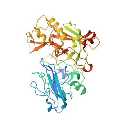
3ZOV

CRYSTAL STRUCTURE OF BACE-1 IN COMPLEX WITH CHEMICAL LIGAND


Experimental Data Snapshot

  • Method: X-RAY DIFFRACTION
  • Resolution: 2.10 Å
  • R-Value Free: 0.272 
  • R-Value Work: 0.219 
  • R-Value Observed: 0.221 

Starting Model: experimental
View more details

wwPDB Validation   3D Report Full Report


Ligand Structure Quality Assessment 


This is version 1.4 of the entry. See complete history


Literature

Mapping the Conformational Space Accessible to Bace2 Using Surface Mutants and Co-Crystals with Fab-Fragments, Fynomers, and Xaperones

Banner, D.W.Gsell, B.Benz, J.Bertschinger, J.Burger, D.Brack, S.Cuppuleri, S.Debulpaep, M.Gast, A.Grabulovski, D.Hennig, M.Hilpert, H.Huber, W.Kuglstatter, A.Kusznir, E.Laeremans, T.Matile, H.Miscenic, C.Rufer, A.Schlatter, D.Steyeart, J.Stihle, M.Thoma, R.Weber, M.Ruf, A.

(2013) Acta Crystallogr D Biol Crystallogr 69: 1124

  • DOI: https://doi.org/10.1107/S0907444913006574
  • Primary Citation of Related Structures:  
    3ZKG, 3ZKI, 3ZKM, 3ZKN, 3ZKQ, 3ZKS, 3ZKX, 3ZL7, 3ZOV, 4BEL, 4BFB

  • PubMed Abstract: 

    The aspartic protease BACE2 is responsible for the shedding of the transmembrane protein Tmem27 from the surface of pancreatic β-cells, which leads to inactivation of the β-cell proliferating activity of Tmem27. This role of BACE2 in the control of β-cell maintenance suggests BACE2 as a drug target for diabetes. Inhibition of BACE2 has recently been shown to lead to improved control of glucose homeostasis and to increased insulin levels in insulin-resistant mice. BACE2 has 52% sequence identity to the well studied Alzheimer's disease target enzyme β-secretase (BACE1). High-resolution BACE2 structures would contribute significantly to the investigation of this enzyme as either a drug target or anti-target. Surface mutagenesis, BACE2-binding antibody Fab fragments, single-domain camelid antibody VHH fragments (Xaperones) and Fyn-kinase-derived SH3 domains (Fynomers) were used as crystallization helpers to obtain the first high-resolution structures of BACE2. Eight crystal structures in six different packing environments define an ensemble of low-energy conformations available to the enzyme. Here, the different strategies used for raising and selecting BACE2 binders for cocrystallization are described and the crystallization success, crystal quality and the time and resources needed to obtain suitable crystals are compared.


  • Organizational Affiliation

    pRED Pharma Research and Early Development, Small Molecule Research, Discovery Technologies, F. Hoffmann-La Roche Ltd, 4070 Basel, Switzerland.


Macromolecules
Find similar proteins by:  (by identity cutoff)  |  3D Structure
Entity ID: 1
MoleculeChains Sequence LengthOrganismDetailsImage
BETA-SECRETASE 1409Homo sapiensMutation(s): 1 
EC: 3.4.23.46
UniProt & NIH Common Fund Data Resources
Find proteins for P56817 (Homo sapiens)
Explore P56817 
Go to UniProtKB:  P56817
PHAROS:  P56817
GTEx:  ENSG00000186318 
Entity Groups  
Sequence Clusters30% Identity50% Identity70% Identity90% Identity95% Identity100% Identity
UniProt GroupP56817
Sequence Annotations
Expand
  • Reference Sequence
Small Molecules
Ligands 3 Unique
IDChains Name / Formula / InChI Key2D Diagram3D Interactions
WZV
Query on WZV

Download Ideal Coordinates CCD File 
B [auth A]5-(2,2,2-Trifluoro-ethoxy)-pyridine-2-carboxylic acid [3-((S)-2-amino-1,4-dimethyl-6-oxo-1,4,5,6-tetrahydro-pyrimidin-4-yl)-phenyl]-amide
C20 H20 F3 N5 O3
ZJFIBYCNZHIVEL-IBGZPJMESA-N
DMS
Query on DMS

Download Ideal Coordinates CCD File 
E [auth A]DIMETHYL SULFOXIDE
C2 H6 O S
IAZDPXIOMUYVGZ-UHFFFAOYSA-N
NA
Query on NA

Download Ideal Coordinates CCD File 
C [auth A],
D [auth A]
SODIUM ION
Na
FKNQFGJONOIPTF-UHFFFAOYSA-N
Binding Affinity Annotations 
IDSourceBinding Affinity
WZV PDBBind:  3ZOV IC50: 60 (nM) from 1 assay(s)
Experimental Data & Validation

Experimental Data

  • Method: X-RAY DIFFRACTION
  • Resolution: 2.10 Å
  • R-Value Free: 0.272 
  • R-Value Work: 0.219 
  • R-Value Observed: 0.221 
  • Space Group: P 61 2 2
Unit Cell:
Length ( Å )Angle ( ˚ )
a = 102.571α = 90
b = 102.571β = 90
c = 171.32γ = 120
Software Package:
Software NamePurpose
REFMACrefinement
XDSdata reduction
SADABSdata scaling
PHASERphasing

Structure Validation

View Full Validation Report


Ligand Structure Quality Assessment 


Entry History 

Deposition Data

Revision History  (Full details and data files)

  • Version 1.0: 2013-05-29
    Type: Initial release
  • Version 1.1: 2013-06-05
    Changes: Database references
  • Version 1.2: 2019-01-30
    Changes: Data collection, Experimental preparation
  • Version 1.3: 2019-02-06
    Changes: Data collection, Experimental preparation
  • Version 1.4: 2023-12-20
    Changes: Data collection, Database references, Derived calculations, Other, Refinement description