The 'Gating' Residues Ile199 and Tyr326 in Human Monoamine Oxidase B Function in Substrate and Inhibitor Recognition.
Milczek, E.M., Binda, C., Rovida, S., Mattevi, A., Edmondson, D.E.(2011) FEBS J 278: 4860
- PubMed: 21978362 Search on PubMedSearch on PubMed Central
- DOI: https://doi.org/10.1111/j.1742-4658.2011.08386.x
- Primary Citation Related Structures:
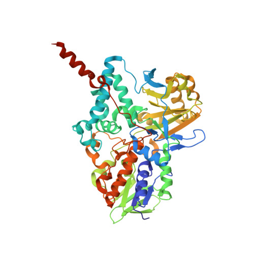
3ZYX - PubMed Abstract:
The major structural difference between human monoamine oxidases A (MAO A) and B (MAO B) is that MAO A has a monopartite substrate cavity of ~550 Å(3) volume and MAO B contains a dipartite cavity structure with volumes of ~290 Å(3) (entrance cavity) and ~400 Å(3) (substrate cavity). Ile199 and Tyr326 side chains separate these two cavities in MAO B. To probe the function of these gating residues, Ile199Ala and Ile199Ala-Tyr326Ala mutant forms of MAO B were investigated. Structural data on the Ile199Ala MAO B mutant show no alterations in active site geometries compared with wild-type enzyme while the Ile199Ala-Tyr326Ala MAO B mutant exhibits alterations in residues 100-103 which are part of the loop gating the entrance to the active site. Both mutant enzymes exhibit catalytic properties with increased amine K(M) but unaltered k(cat) values. The altered K(M) values on mutation are attributed to the influence of the cavity structure in the binding and subsequent deprotonation of the amine substrate. Both mutant enzymes exhibit weaker binding affinities relative to wild-type enzyme for small reversible inhibitors. Ile199Ala MAO B exhibits an increase in binding affinity for reversible MAO B specific inhibitors which bridge both cavities. The Ile199Ala-Tyr326Ala double mutant exhibits inhibitor binding properties more similar to those of MAO A than to MAO B. These results demonstrate that the bipartite cavity structure in MAO B plays an important role in substrate and inhibitor recognition to distinguish its specificities from those of MAO A and provide insights into specific reversible inhibitor design for these membrane-bound enzymes.
- Department of Chemistry, Emory University, Atlanta, Georgia 30322, USA.
Organizational Affiliation: