3CSB | pdb_00003csb

Crystal Structure of Monobody YSX1/Maltose Binding Protein Fusion Complex


Experimental Data Snapshot

  • Method: X-RAY DIFFRACTION
  • Resolution: 2.00 Å
  • R-Value Free: 
    0.236 (Depositor), 0.229 (DCC) 
  • R-Value Work: 
    0.195 (Depositor), 0.193 (DCC) 
  • R-Value Observed: 
    0.199 (Depositor) 

wwPDB Validation 3D Report Full Report

Validation slider image for 3CSB

This is version 1.4 of the entry. See complete history

Literature

A Dominant Conformational Role for Amino Acid Diversity in Minimalist Protein-Protein Interfaces

Gilbreth, R.N.Esaki, K.Koide, A.Sidhu, S.S.Koide, S.

(2008) J Mol Biology 381: 407-418

  • DOI: https://doi.org/10.1016/j.jmb.2008.06.014
  • Primary Citation Related Structures: 
    3CSB, 3CSG

  • PubMed Abstract: 

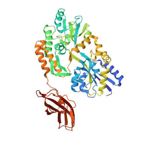
    Recent studies have shown that highly simplified interaction surfaces consisting of combinations of just two amino acids, Tyr and Ser, exhibit high affinity and specificity. The high functional levels of such minimalist interfaces might thus indicate small contributions of greater amino acid diversity seen in natural interfaces. Toward addressing this issue, we have produced a pair of binding proteins built on the fibronectin type III scaffold, termed "monobodies." One monobody contains the Tyr/Ser binary-code interface (termed YS) and the other contains an expanded amino acid diversity interface (YSX), but both bind to an identical target, maltose-binding protein. The YSX monobody bound with higher affinity, a slower off rate and a more favorable enthalpic contribution than the YS monobody. High-resolution X-ray crystal structures revealed that both proteins bound to an essentially identical epitope, providing a unique opportunity to directly investigate the role of amino acid diversity in a protein interaction interface. Surprisingly, Tyr still dominates the YSX paratope and the additional amino acid types are primarily used to conformationally optimize contacts made by tyrosines. Scanning mutagenesis showed that while all contacting Tyr side chains are essential in the YS monobody, the YSX interface was more tolerant to mutations. These results suggest that the conformational, not chemical, diversity of additional types of amino acids provided higher functionality and evolutionary robustness, supporting the dominant role of Tyr and the importance of conformational diversity in forming protein interaction interfaces.


  • Organizational Affiliation
    • Department of Biochemistry and Molecular Biology, The University of Chicago, 929 East 57th Street, Chicago, IL 60637, USA.

Macromolecule Content 

  • Total Structure Weight: 52.66 kDa 
  • Atom Count: 3,929 
  • Modeled Residue Count: 464 
  • Deposited Residue Count: 465 
  • Unique protein chains: 1

Macromolecules

Find similar proteins by:|  3D Structure
Entity ID: 1
MoleculeChains  Sequence LengthOrganismDetailsImage
Maltose-binding protein Monobody YSX1 Fusion465N/AMutation(s): 0 
Gene Names: malE
UniProt
Find proteins for P0AEX9 (Escherichia coli (strain K12))
Explore P0AEX9 
Go to UniProtKB:  P0AEX9
Entity Groups
Sequence Clusters30% Identity50% Identity70% Identity90% Identity95% Identity100% Identity
UniProt GroupP0AEX9
Sequence Annotations
Expand
Reference Sequence

Small Molecules

Ligands 6 Unique
IDChains Name / Formula / InChI Key2D Diagram3D Interactions
1PE

Query on 1PE


Download:Ideal Coordinates CCD File
J [auth A]PENTAETHYLENE GLYCOL
C10 H22 O6
JLFNLZLINWHATN-UHFFFAOYSA-N
PG4

Query on PG4


Download:Ideal Coordinates CCD File
I [auth A]TETRAETHYLENE GLYCOL
C8 H18 O5
UWHCKJMYHZGTIT-UHFFFAOYSA-N
PGE

Query on PGE


Download:Ideal Coordinates CCD File
H [auth A]TRIETHYLENE GLYCOL
C6 H14 O4
ZIBGPFATKBEMQZ-UHFFFAOYSA-N
PEG

Query on PEG


Download:Ideal Coordinates CCD File
K [auth A]
L [auth A]
M [auth A]
N [auth A]
O [auth A]
K [auth A],
L [auth A],
M [auth A],
N [auth A],
O [auth A],
P [auth A],
Q [auth A],
R [auth A],
S [auth A]
DI(HYDROXYETHYL)ETHER
C4 H10 O3
MTHSVFCYNBDYFN-UHFFFAOYSA-N
EDO

Query on EDO


Download:Ideal Coordinates CCD File
F [auth A],
G [auth A]
1,2-ETHANEDIOL
C2 H6 O2
LYCAIKOWRPUZTN-UHFFFAOYSA-N
MN

Query on MN


Download:Ideal Coordinates CCD File
B [auth A],
C [auth A],
D [auth A],
E [auth A]
MANGANESE (II) ION
Mn
WAEMQWOKJMHJLA-UHFFFAOYSA-N

Experimental Data & Validation

Experimental Data

  • Method: X-RAY DIFFRACTION
  • Resolution: 2.00 Å
  • R-Value Free:  0.236 (Depositor), 0.229 (DCC) 
  • R-Value Work:  0.195 (Depositor), 0.193 (DCC) 
  • R-Value Observed: 0.199 (Depositor) 
Space Group: P 41 21 2
Unit Cell:
Length ( Å )Angle ( ˚ )
a = 98.818α = 90
b = 98.818β = 90
c = 134.619γ = 90
Software Package:
Software NamePurpose
REFMACrefinement
CNSrefinement
Blu-Icedata collection
HKL-2000data reduction
HKL-2000data scaling
CNSphasing
REFMACphasing

Structure Validation

View Full Validation Report


Entry History 

Deposition Data

Revision History  (Full details and data files)

  • Version 1.0: 2008-07-22
    Type: Initial release
  • Version 1.1: 2011-07-13
    Changes: Version format compliance
  • Version 1.2: 2017-10-25
    Changes: Refinement description
  • Version 1.3: 2019-07-24
    Changes: Data collection, Refinement description
  • Version 1.4: 2024-02-21
    Changes: Data collection, Database references, Derived calculations