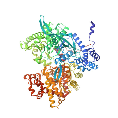
3E3O

Glycogen phosphorylase R state-IMP complex


Experimental Data Snapshot

  • Method: X-RAY DIFFRACTION
  • Resolution: 2.60 Å
  • R-Value Free: 0.267 
  • R-Value Work: 0.201 
  • R-Value Observed: 0.204 

Starting Model: experimental
View more details

wwPDB Validation   3D Report Full Report


Ligand Structure Quality Assessment 


This is version 1.5 of the entry. See complete history


Literature

Glycogen phosphorylase revisited: extending the resolution of the R- and T-state structures of the free enzyme and in complex with allosteric activators.

Leonidas, D.D.Zographos, S.E.Tsitsanou, K.E.Skamnaki, V.T.Stravodimos, G.Kyriakis, E.

(2021) Acta Crystallogr F Struct Biol Commun 77: 303-311

  • DOI: https://doi.org/10.1107/S2053230X21008542
  • Primary Citation of Related Structures:  
    3E3L, 3E3N, 3E3O, 7P7D

  • PubMed Abstract: 

    The crystal structures of free T-state and R-state glycogen phosphorylase (GP) and of R-state GP in complex with the allosteric activators IMP and AMP are reported at improved resolution. GP is a validated pharmaceutical target for the development of antihyperglycaemic agents, and the reported structures may have a significant impact on structure-based drug-design efforts. Comparisons with previously reported structures at lower resolution reveal the detailed conformation of important structural features in the allosteric transition of GP from the T-state to the R-state. The conformation of the N-terminal segment (residues 7-17), the position of which was not located in previous T-state structures, was revealed to form an α-helix (now termed α0). The conformation of this segment (which contains Ser14, phosphorylation of which leads to the activation of GP) is significantly different between the T-state and the R-state, pointing in opposite directions. In the T-state it is packed between helices α4 and α16 (residues 104-115 and 497-508, respectively), while in the R-state it is packed against helix α1 (residues 22'-38') and towards the loop connecting helices α4' and α5' of the neighbouring subunit. The allosteric binding site where AMP and IMP bind is formed by the ordering of a loop (residues 313-326) which is disordered in the free structure, and adopts a conformation dictated mainly by the type of nucleotide that binds at this site.


  • Organizational Affiliation

    Department of Biochemistry and Biotechnology, University of Thessaly, Biopolis, 41500 Larissa, Greece.


Macromolecules
Find similar proteins by:  (by identity cutoff)  |  3D Structure
Entity ID: 1
MoleculeChains Sequence LengthOrganismDetailsImage
Glycogen phosphorylase, muscle form
A, B, C, D
842Oryctolagus cuniculusMutation(s): 0 
EC: 2.4.1.1
UniProt
Find proteins for P00489 (Oryctolagus cuniculus)
Explore P00489 
Go to UniProtKB:  P00489
Entity Groups  
Sequence Clusters30% Identity50% Identity70% Identity90% Identity95% Identity100% Identity
UniProt GroupP00489
Sequence Annotations
Expand
  • Reference Sequence
Small Molecules
Ligands 2 Unique
IDChains Name / Formula / InChI Key2D Diagram3D Interactions
IMP
Query on IMP

Download Ideal Coordinates CCD File 
F [auth A],
J [auth B],
M [auth C],
P [auth D]
INOSINIC ACID
C10 H13 N4 O8 P
GRSZFWQUAKGDAV-KQYNXXCUSA-N
SO4
Query on SO4

Download Ideal Coordinates CCD File 
E [auth A]
G [auth B]
H [auth B]
I [auth B]
K [auth C]
E [auth A],
G [auth B],
H [auth B],
I [auth B],
K [auth C],
L [auth C],
N [auth D],
O [auth D]
SULFATE ION
O4 S
QAOWNCQODCNURD-UHFFFAOYSA-L
Modified Residues  1 Unique
IDChains TypeFormula2D DiagramParent
LLP
Query on LLP
A, B, C, D
L-PEPTIDE LINKINGC14 H22 N3 O7 PLYS
Experimental Data & Validation

Experimental Data

  • Method: X-RAY DIFFRACTION
  • Resolution: 2.60 Å
  • R-Value Free: 0.267 
  • R-Value Work: 0.201 
  • R-Value Observed: 0.204 
  • Space Group: P 1 21 1
Unit Cell:
Length ( Å )Angle ( ˚ )
a = 118.68α = 90
b = 188.448β = 109.1
c = 87.853γ = 90
Software Package:
Software NamePurpose
REFMACrefinement
MAR345data collection
DENZOdata reduction
SCALEPACKdata scaling
REFMACphasing

Structure Validation

View Full Validation Report


Ligand Structure Quality Assessment 


Entry History 

Deposition Data

Revision History  (Full details and data files)

  • Version 1.0: 2009-08-18
    Type: Initial release
  • Version 1.1: 2011-07-13
    Changes: Advisory, Version format compliance
  • Version 1.2: 2017-10-25
    Changes: Refinement description
  • Version 1.3: 2021-09-15
    Changes: Data collection, Database references, Derived calculations
  • Version 1.4: 2023-11-01
    Changes: Data collection, Refinement description
  • Version 1.5: 2023-11-15
    Changes: Data collection