


Crystal structure of the 1E9 PheL89Ser/LeuH47Trp/MetH100bPhe Fab in complex with a 39A11 transition state analog
Verdino, P., Aldag, C., Jonsson, S., Hilvert, D., Wilson, I.A.To be published.
Experimental Data Snapshot
Starting Model: experimental
View more details
Entity ID: 1 | |||||
|---|---|---|---|---|---|
| Molecule | Chains | Sequence Length | Organism | Details | Image |
| Chimeric antibody Fab 1E9, light chain | A [auth L] | 219 | Mus musculus, Homo sapiens This entity is chimeric | Mutation(s): 0 | 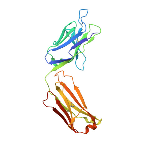 |
Entity ID: 2 | |||||
|---|---|---|---|---|---|
| Molecule | Chains | Sequence Length | Organism | Details | Image |
| Chimeric antibody Fab 1E9, heavy chain | B [auth H] | 227 | Mus musculus, Homo sapiens This entity is chimeric | Mutation(s): 0 | 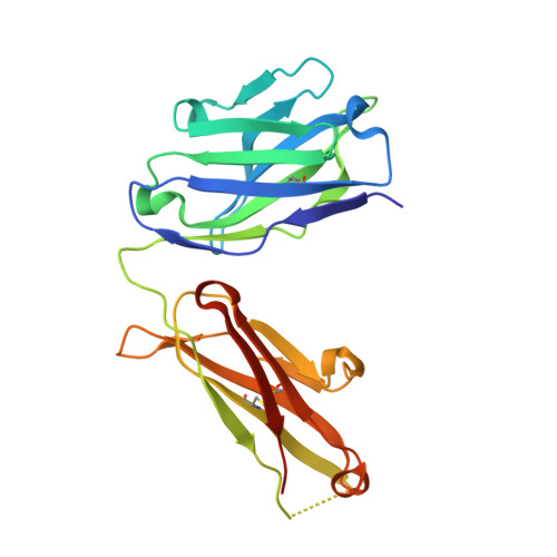 |
| Ligands 4 Unique | |||||
|---|---|---|---|---|---|
| ID | Chains | Name / Formula / InChI Key | 2D Diagram | 3D Interactions | |
| O2W Download:Ideal Coordinates CCD File | C [auth L] | methyl ({[(3aR,4R,7R,7aR)-2-(4-aminophenyl)-1,3-dioxooctahydro-4H-4,7-ethanoisoindol-4-yl]carbamoyl}oxy)acetate C20 H23 N3 O6 PWUNEFYJETTWLK-KCQBUTHOSA-N |  | ||
| FLC Download:Ideal Coordinates CCD File | S [auth H], T [auth H], U [auth H] | CITRATE ANION C6 H5 O7 KRKNYBCHXYNGOX-UHFFFAOYSA-K |  | ||
| TRS Download:Ideal Coordinates CCD File | D [auth L] E [auth L] F [auth L] G [auth L] N [auth H] | 2-AMINO-2-HYDROXYMETHYL-PROPANE-1,3-DIOL C4 H12 N O3 LENZDBCJOHFCAS-UHFFFAOYSA-O |  | ||
| SO4 Download:Ideal Coordinates CCD File | H [auth L] I [auth L] J [auth L] K [auth L] L | SULFATE ION O4 S QAOWNCQODCNURD-UHFFFAOYSA-L |  | ||
| Length ( Å ) | Angle ( ˚ ) |
|---|---|
| a = 128.203 | α = 90 |
| b = 128.203 | β = 90 |
| c = 92.144 | γ = 120 |
| Software Name | Purpose |
|---|---|
| BOS | data collection |
| REFMAC | refinement |
| HKL-2000 | data reduction |
| HKL-2000 | data scaling |
| REFMAC | phasing |


