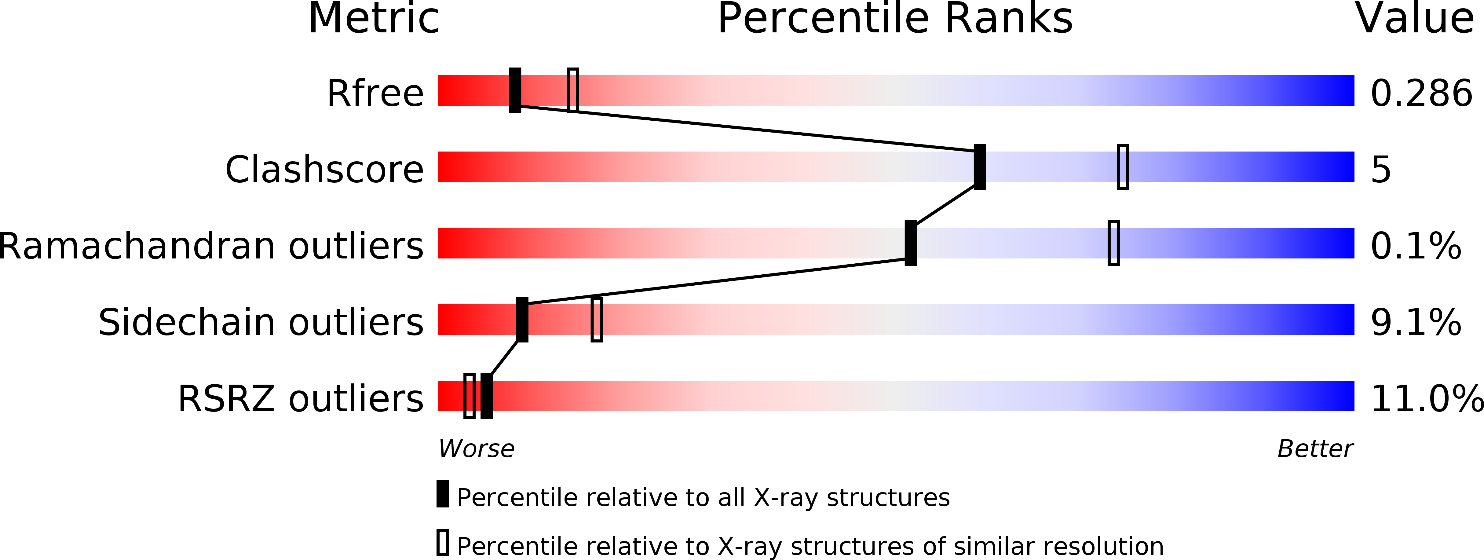
Structures of AzrA and of AzrC complexed with substrate or inhibitor: insight into substrate specificity and catalytic mechanism.
Yu, J., Ogata, D., Gai, Z., Taguchi, S., Tanaka, I., Ooi, T., Yao, M.(2014) Acta Crystallogr D Biol Crystallogr 70: 553-564
- PubMed: 24531489
- DOI: https://doi.org/10.1107/S1399004713030988
- Primary Citation of Related Structures:
3W77, 3W78, 3W79, 3W7A - PubMed Abstract:
Azo dyes are major synthetic dyestuffs with one or more azo bonds and are widely used for various industrial purposes. The biodegradation of residual azo dyes via azoreductase-catalyzed cleavage is very efficient as the initial step of wastewater treatment. The structures of the complexes of azoreductases with various substrates are therefore indispensable to understand their substrate specificity and catalytic mechanism. In this study, the crystal structures of AzrA and of AzrC complexed with Cibacron Blue (CB) and the azo dyes Acid Red 88 (AR88) and Orange I (OI) were determined. As an inhibitor/analogue of NAD(P)H, CB was located on top of flavin mononucleotide (FMN), suggesting a similar binding manner as NAD(P)H for direct hydride transfer to FMN. The structures of the AzrC-AR88 and AzrC-OI complexes showed two manners of binding for substrates possessing a hydroxy group at the ortho or the para position of the azo bond, respectively, while AR88 and OI were estimated to have a similar binding affinity to AzrC from ITC experiments. Although the two substrates were bound in different orientations, the hydroxy groups were located in similar positions, resulting in an arrangement of electrophilic C atoms binding with a proton/electron-donor distance of ∼3.5 Å to N5 of FMN. Catalytic mechanisms for different substrates are proposed based on the crystal structures and on site-directed mutagenesis analysis.
Organizational Affiliation:
Faculty of Advanced Life Science, Hokkaido University, Kita 10, Nishi 8, Kita-ku, Sapporo, Hokkaido 060-0810, Japan.


