4ACB | pdb_00004acb

CRYSTAL STRUCTURE OF TRANSLATION ELONGATION FACTOR SELB FROM METHANOCOCCUS MARIPALUDIS IN COMPLEX WITH THE GTP ANALOGUE GPPNHP


Experimental Data Snapshot

  • Method: X-RAY DIFFRACTION
  • Resolution: 3.34 Å
  • R-Value Free: 
    0.223 (Depositor), 0.230 (DCC) 
  • R-Value Work: 
    0.179 (Depositor), 0.189 (DCC) 
  • R-Value Observed: 
    0.181 (Depositor) 

Starting Model: experimental
View more details

wwPDB Validation 3D Report Full Report

Validation slider image for 4ACB

Ligand Structure Quality Assessment 


This is version 1.2 of the entry. See complete history

Literature

Selenocysteine tRNA-Specific Elongation Factor Selb is a Structural Chimaera of Elongation and Initiation Factors.

Leibundgut, M.Frick, C.Thanbichler, M.Bock, A.Ban, N.

(2005) EMBO J 24: 11

  • DOI: https://doi.org/10.1038/sj.emboj.7600505
  • Primary Citation Related Structures: 
    4AC9, 4ACA, 4ACB

  • PubMed Abstract: 

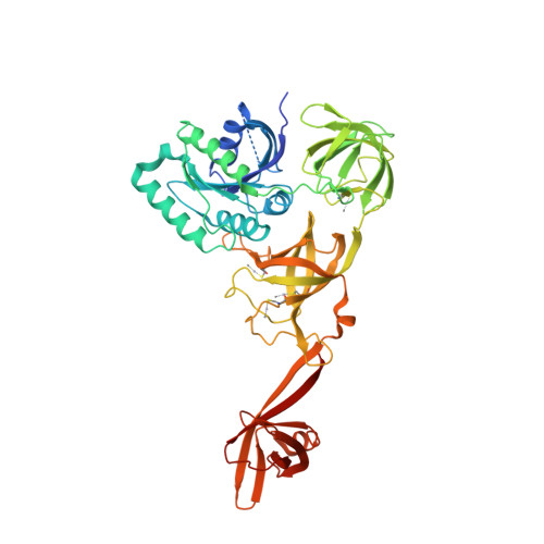
    In all three kingdoms of life, SelB is a specialized translation elongation factor responsible for the cotranslational incorporation of selenocysteine into proteins by recoding of a UGA stop codon in the presence of a downstream mRNA hairpin loop. Here, we present the X-ray structures of SelB from the archaeon Methanococcus maripaludis in the apo-, GDP- and GppNHp-bound form and use mutational analysis to investigate the role of individual amino acids in its aminoacyl-binding pocket. All three SelB structures reveal an EF-Tu:GTP-like domain arrangement. Upon binding of the GTP analogue GppNHp, a conformational change of the Switch 2 region in the GTPase domain leads to the exposure of SelB residues involved in clamping the 5' phosphate of the tRNA. A conserved extended loop in domain III of SelB may be responsible for specific interactions with tRNA(Sec) and act as a ruler for measuring the extra long acceptor arm. Domain IV of SelB adopts a beta barrel fold and is flexibly tethered to domain III. The overall domain arrangement of SelB resembles a 'chalice' observed so far only for initiation factor IF2/eIF5B. In our model of SelB bound to the ribosome, domain IV points towards the 3' mRNA entrance cleft ready to interact with the downstream secondary structure element.


  • Organizational Affiliation
    • Institut für Molekularbiologie und Biophysik, Eidgenössische Technische Hochschule Zürich, Zürich, Switzerland.

Macromolecule Content 

  • Total Structure Weight: 221.29 kDa 
  • Atom Count: 14,623 
  • Modeled Residue Count: 1,846 
  • Deposited Residue Count: 1,928 
  • Unique protein chains: 1

Macromolecules

Find similar proteins by:|  3D Structure
Entity ID: 1
MoleculeChains  Sequence LengthOrganismDetailsImage
TRANSLATION ELONGATION FACTOR SELB
A, B, C, D
482Methanococcus maripaludisMutation(s): 0 
UniProt
Find proteins for Q8J307 (Methanococcus maripaludis)
Explore Q8J307 
Go to UniProtKB:  Q8J307
Entity Groups
Sequence Clusters30% Identity50% Identity70% Identity90% Identity95% Identity100% Identity
UniProt GroupQ8J307
Sequence Annotations
Expand
Reference Sequence

Small Molecules

Ligands 6 Unique
IDChains Name / Formula / InChI Key2D Diagram3D Interactions
GNP

Query on GNP


Download:Ideal Coordinates CCD File
E [auth A]PHOSPHOAMINOPHOSPHONIC ACID-GUANYLATE ESTER
C10 H17 N6 O13 P3
UQABYHGXWYXDTK-UUOKFMHZSA-N
GDP

Query on GDP


Download:Ideal Coordinates CCD File
G [auth B]GUANOSINE-5'-DIPHOSPHATE
C10 H15 N5 O11 P2
QGWNDRXFNXRZMB-UUOKFMHZSA-N
DXC

Query on DXC


Download:Ideal Coordinates CCD File
K [auth B]
P [auth C]
Q [auth C]
R [auth C]
S [auth C]
K [auth B],
P [auth C],
Q [auth C],
R [auth C],
S [auth C],
T [auth C],
U [auth C]
(3ALPHA,5BETA,12ALPHA)-3,12-DIHYDROXYCHOLAN-24-OIC ACID
C24 H40 O4
KXGVEGMKQFWNSR-LLQZFEROSA-N
5GP

Query on 5GP


Download:Ideal Coordinates CCD File
L [auth B]GUANOSINE-5'-MONOPHOSPHATE
C10 H14 N5 O8 P
RQFCJASXJCIDSX-UUOKFMHZSA-N
SO4

Query on SO4


Download:Ideal Coordinates CCD File
I [auth B]
J [auth B]
M [auth C]
N [auth C]
O [auth C]
I [auth B],
J [auth B],
M [auth C],
N [auth C],
O [auth C],
V [auth C]
SULFATE ION
O4 S
QAOWNCQODCNURD-UHFFFAOYSA-L
MG

Query on MG


Download:Ideal Coordinates CCD File
F [auth A],
H [auth B]
MAGNESIUM ION
Mg
JLVVSXFLKOJNIY-UHFFFAOYSA-N
Modified Residues  1 Unique
IDChains TypeFormula2D DiagramParent
CMH
Query on CMH
A, B, C, D
L-PEPTIDE LINKINGC4 H9 Hg N O2 SCYS

Experimental Data & Validation

Experimental Data

  • Method: X-RAY DIFFRACTION
  • Resolution: 3.34 Å
  • R-Value Free:  0.223 (Depositor), 0.230 (DCC) 
  • R-Value Work:  0.179 (Depositor), 0.189 (DCC) 
  • R-Value Observed: 0.181 (Depositor) 
Space Group: P 31 1 2
Unit Cell:
Length ( Å )Angle ( ˚ )
a = 146.63α = 90
b = 146.63β = 90
c = 297.22γ = 120
Software Package:
Software NamePurpose
PHENIXrefinement
XDSdata reduction
XSCALEdata scaling

Structure Validation

View Full Validation Report


Ligand Structure Quality Assessment 


Entry History 

Deposition Data

Revision History  (Full details and data files)

  • Version 1.0: 2012-11-07
    Type: Initial release
  • Version 1.1: 2023-12-20
    Changes: Data collection, Database references, Derived calculations, Other, Refinement description
  • Version 1.2: 2024-10-23
    Changes: Structure summary