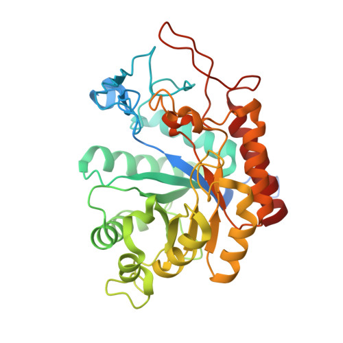
4AD4

Structure of the GH99 endo-alpha-mannosidase from Bacteroides xylanisolvens in complex with glucose-1,3-isofagomine and alpha-1,2- mannobiose


Experimental Data Snapshot

  • Method: X-RAY DIFFRACTION
  • Resolution: 1.90 Å
  • R-Value Free: 0.190 
  • R-Value Work: 0.156 
  • R-Value Observed: 0.158 

Starting Model: experimental
View more details

wwPDB Validation   3D Report Full Report


Ligand Structure Quality Assessment 


This is version 2.1 of the entry. See complete history


Literature

Structural and Mechanistic Insight Into N-Glycan Processing by Endo-Alpha-Mannosidase.

Thompson, A.J.Williams, R.J.Hakki, Z.Alonzi, D.S.Wennekes, T.Gloster, T.M.Songsrirote, K.Thomas-Oates, J.E.Wrodnigg, T.M.Spreitz, J.Stutz, A.E.Butters, T.D.Williams, S.J.Davies, G.J.

(2012) Proc Natl Acad Sci U S A 109: 781

  • DOI: https://doi.org/10.1073/pnas.1111482109
  • Primary Citation of Related Structures:  
    4ACY, 4ACZ, 4AD0, 4AD1, 4AD2, 4AD3, 4AD4, 4AD5

  • PubMed Abstract: 

    N-linked glycans play key roles in protein folding, stability, and function. Biosynthetic modification of N-linked glycans, within the endoplasmic reticulum, features sequential trimming and readornment steps. One unusual enzyme, endo-α-mannosidase, cleaves mannoside linkages internally within an N-linked glycan chain, short circuiting the classical N-glycan biosynthetic pathway. Here, using two bacterial orthologs, we present the first structural and mechanistic dissection of endo-α-mannosidase. Structures solved at resolutions 1.7-2.1 Å reveal a (β/α)(8) barrel fold in which the catalytic center is present in a long substrate-binding groove, consistent with cleavage within the N-glycan chain. Enzymatic cleavage of authentic Glc(1/3)Man(9)GlcNAc(2) yields Glc(1/3)-Man. Using the bespoke substrate α-Glc-1,3-α-Man fluoride, the enzyme was shown to act with retention of anomeric configuration. Complexes with the established endo-α-mannosidase inhibitor α-Glc-1,3-deoxymannonojirimycin and a newly developed inhibitor, α-Glc-1,3-isofagomine, and with the reducing-end product α-1,2-mannobiose structurally define the -2 to +2 subsites of the enzyme. These structural and mechanistic data provide a foundation upon which to develop new enzyme inhibitors targeting the hijacking of N-glycan synthesis in viral disease and cancer.


  • Organizational Affiliation

    Department of Chemistry, University of York, Heslington, York YO10 5DD, United Kingdom.


Macromolecules
Find similar proteins by:  (by identity cutoff)  |  3D Structure
Entity ID: 1
MoleculeChains Sequence LengthOrganismDetailsImage
GLYCOSYL HYDROLASE FAMILY 71380Bacteroides xylanisolvens XB1AMutation(s): 0 
EC: 3.2.1.130
UniProt
Find proteins for D6D1V7 (Bacteroides xylanisolvens XB1A)
Explore D6D1V7 
Go to UniProtKB:  D6D1V7
Entity Groups  
Sequence Clusters30% Identity50% Identity70% Identity90% Identity95% Identity100% Identity
UniProt GroupD6D1V7
Sequence Annotations
Expand
  • Reference Sequence
Oligosaccharides

Help

Entity ID: 2
MoleculeChains Length2D Diagram Glycosylation3D Interactions
alpha-D-mannopyranose-(1-2)-alpha-D-mannopyranose
B
2N/A
Glycosylation Resources
GlyTouCan:  G53402KW
GlyCosmos:  G53402KW
GlyGen:  G53402KW
Small Molecules
Biologically Interesting Molecules (External Reference) 1 Unique
Experimental Data & Validation

Experimental Data

  • Method: X-RAY DIFFRACTION
  • Resolution: 1.90 Å
  • R-Value Free: 0.190 
  • R-Value Work: 0.156 
  • R-Value Observed: 0.158 
  • Space Group: I 4
Unit Cell:
Length ( Å )Angle ( ˚ )
a = 108.37α = 90
b = 108.37β = 90
c = 67.65γ = 90
Software Package:
Software NamePurpose
REFMACrefinement
MOSFLMdata reduction
SCALAdata scaling
MOLREPphasing

Structure Validation

View Full Validation Report


Ligand Structure Quality Assessment 


Entry History 

Revision History  (Full details and data files)

  • Version 1.0: 2012-02-01
    Type: Initial release
  • Version 1.1: 2012-02-29
    Changes: Other
  • Version 1.2: 2012-04-18
    Changes: Other
  • Version 1.3: 2015-07-15
    Changes: Data collection, Non-polymer description
  • Version 2.0: 2020-07-29
    Type: Remediation
    Reason: Carbohydrate remediation
    Changes: Advisory, Atomic model, Data collection, Derived calculations, Other, Structure summary
  • Version 2.1: 2024-05-01
    Changes: Data collection, Database references, Refinement description, Structure summary