Characterization of a Small Molecule Inhibitor of Plasminogen Activator Inhibitor Type 1 that Accelerates the Transition Into the Latent Conformation
Fjellstrom, O., Deinum, J., Sjogren, T., Johansson, C., Geschwindner, S., Nerme, V., Legnehed, A., Mcpheat, J., Olsson, K., Bodin, C., Panuovic, A., Gustafsson, D.(2013) J Biol Chem 288: 873
- PubMed: 23155046 Search on PubMedSearch on PubMed Central
- DOI: https://doi.org/10.1074/jbc.M112.371732
- Primary Citation Related Structures:
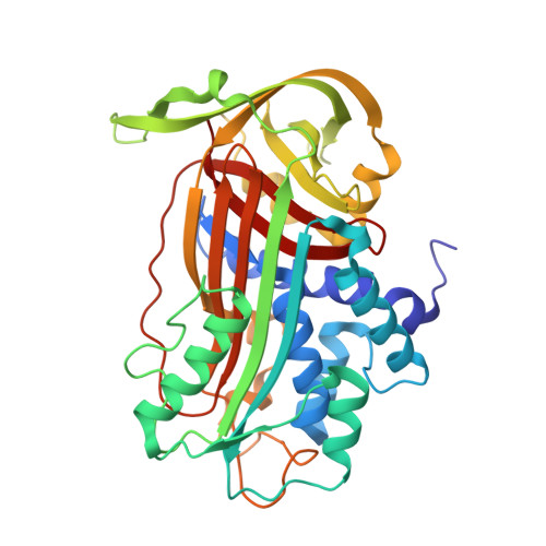
4AQH - PubMed Abstract:
A novel class of small molecule inhibitors for plasminogen activator inhibitor type 1 (PAI-1), represented by AZ3976, was identified in a high throughput screening campaign. AZ3976 displayed an IC(50) value of 26 μm in an enzymatic chromogenic assay. In a plasma clot lysis assay, the compound was active with an IC(50) of 16 μm. Surprisingly, AZ3976 did not bind to active PAI-1 but bound to latent PAI-1 with a K(D) of 0.29 μm at 35 °C and a binding stoichiometry of 0.94, as measured by isothermal calorimetry. Reversible binding was confirmed by surface plasmon resonance direct binding experiments. The x-ray structure of AZ3976 in complex with latent PAI-1 was determined at 2.4 Å resolution. The inhibitor was bound in the flexible joint region with the entrance to the cavity located between α-helix D and β-strand 2A. A set of surface plasmon resonance experiments revealed that AZ3976 inhibited PAI-1 by enhancing the latency transition of active PAI-1. Because AZ3976 only had measurable affinity for latent PAI-1, we propose that its mechanism of inhibition is based on binding to a small fraction in equilibrium with active PAI-1, a latent-like prelatent form, from which latent PAI-1 is then generated more rapidly. This mode of action, with induced accelerated latency transition of active PAI-1 may, together with supporting x-ray data, provide improved opportunities for small molecule drug design in the hunt for therapeutically useful PAI-1 inhibitors.
- Department of Medicinal Chemistry, AstraZeneca R&D Mölndal, S-431 83 Mölndal, Sweden. ola.fjellstrom@astrazeneca.com
Organizational Affiliation: