4AW0 | pdb_00004aw0

Human PDK1 Kinase Domain in Complex with Allosteric Compound PS182 Bound to the PIF-Pocket


Experimental Data Snapshot

  • Method: X-RAY DIFFRACTION
  • Resolution: 1.43 Å
  • R-Value Free: 
    0.188 (Depositor), 0.183 (DCC) 
  • R-Value Work: 
    0.170 (Depositor), 0.167 (DCC) 
  • R-Value Observed: 
    0.171 (Depositor) 

Starting Model: experimental
View more details

wwPDB Validation 3D Report Full Report

Validation slider image for 4AW0

Ligand Structure Quality Assessment 


This is version 2.1 of the entry. See complete history

Literature

Substrate-Selective Inhibition of Protein Kinase Pdk1 by Small Compounds that Bind to the Pif-Pocket Allosteric Docking Site.

Busschots, K.Lopez-Garcia, L.A.Lammi, C.Stroba, A.Zeuzem, S.Piiper, A.Alzari, P.M.Neimanis, S.Arencibia, J.M.Engel, M.Schulze, J.O.Biondi, R.M.

(2012) Chem Biol 19: 1152

  • DOI: https://doi.org/10.1016/j.chembiol.2012.07.017
  • Primary Citation Related Structures: 
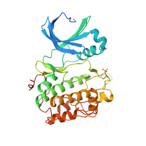
    4AW0, 4AW1

  • PubMed Abstract: 

    The PIF-pocket of AGC protein kinases participates in the physiologic mechanism of regulation by acting as a docking site for substrates and as a switch for the transduction of the conformational changes needed for activation or inhibition. We describe the effects of compounds that bind to the PIF-pocket of PDK1. In vitro, PS210 is a potent activator of PDK1, and the crystal structure of the PDK1-ATP-PS210 complex shows that PS210 stimulates the closure of the kinase domain. However, in cells, the prodrug of PS210 (PS423) acts as a substrate-selective inhibitor of PDK1, inhibiting the phosphorylation and activation of S6K, which requires docking to the PIF-pocket, but not affecting PKB/Akt. This work describes a tool to study the dynamics of PDK1 activity and a potential approach for drug discovery.


  • Organizational Affiliation
    • Research Group PhosphoSites, Department of Internal Medicine I, Universitätsklinikum Frankfurt, Theodor-Stern-Kai 7, 60590 Frankfurt, Germany.

Macromolecule Content 

  • Total Structure Weight: 36.6 kDa 
  • Atom Count: 2,798 
  • Modeled Residue Count: 285 
  • Deposited Residue Count: 311 
  • Unique protein chains: 1

Macromolecules

Find similar proteins by:|  3D Structure
Entity ID: 1
MoleculeChains  Sequence LengthOrganismDetailsImage
3-PHOSPHOINOSITIDE-DEPENDENT PROTEIN KINASE 1311Homo sapiensMutation(s): 2 
EC: 2.7.11.1
UniProt & NIH Common Fund Data Resources
Find proteins for O15530 (Homo sapiens)
Explore O15530 
Go to UniProtKB:  O15530
PHAROS:  O15530
GTEx:  ENSG00000140992 
Entity Groups
Sequence Clusters30% Identity50% Identity70% Identity90% Identity95% Identity100% Identity
UniProt GroupO15530
Sequence Annotations
Expand
Reference Sequence

Small Molecules

Ligands 5 Unique
IDChains Name / Formula / InChI Key2D Diagram3D Interactions
ATP

Query on ATP


Download:Ideal Coordinates CCD File
B [auth A]ADENOSINE-5'-TRIPHOSPHATE
C10 H16 N5 O13 P3
ZKHQWZAMYRWXGA-KQYNXXCUSA-N
MJF

Query on MJF


Download:Ideal Coordinates CCD File
C [auth A][(1R)-3-(4-chlorophenyl)-3-oxo-1-phenylpropyl]propanedioic acid
C18 H15 Cl O5
XOOQYQRTMFAOAP-AWEZNQCLSA-N
DTD

Query on DTD


Download:Ideal Coordinates CCD File
G [auth A]DITHIANE DIOL
C4 H8 O2 S2
YPGMOWHXEQDBBV-IMJSIDKUSA-N
DMS

Query on DMS


Download:Ideal Coordinates CCD File
D [auth A]DIMETHYL SULFOXIDE
C2 H6 O S
IAZDPXIOMUYVGZ-UHFFFAOYSA-N
MG

Query on MG


Download:Ideal Coordinates CCD File
E [auth A],
F [auth A]
MAGNESIUM ION
Mg
JLVVSXFLKOJNIY-UHFFFAOYSA-N
Modified Residues  2 Unique
IDChains TypeFormula2D DiagramParent
CME
Query on CME
A
L-PEPTIDE LINKINGC5 H11 N O3 S2CYS
SEP
Query on SEP
A
L-PEPTIDE LINKINGC3 H8 N O6 PSER

Experimental Data & Validation

Experimental Data

  • Method: X-RAY DIFFRACTION
  • Resolution: 1.43 Å
  • R-Value Free:  0.188 (Depositor), 0.183 (DCC) 
  • R-Value Work:  0.170 (Depositor), 0.167 (DCC) 
  • R-Value Observed: 0.171 (Depositor) 
Space Group: C 1 2 1
Unit Cell:
Length ( Å )Angle ( ˚ )
a = 148.27α = 90
b = 44.01β = 101.57
c = 47.39γ = 90
Software Package:
Software NamePurpose
PHENIXrefinement
XDSdata reduction
XSCALEdata scaling
PHASERphasing

Structure Validation

View Full Validation Report


Ligand Structure Quality Assessment 


Entry History 

Revision History  (Full details and data files)

  • Version 1.0: 2012-10-03
    Type: Initial release
  • Version 1.1: 2012-10-10
    Changes: Database references, Derived calculations, Other, Structure summary
  • Version 2.0: 2023-12-20
    Changes: Atomic model, Data collection, Database references, Derived calculations, Other, Refinement description
  • Version 2.1: 2024-11-13
    Changes: Structure summary