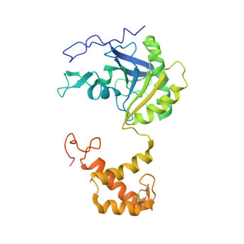
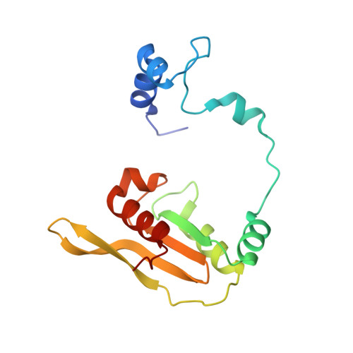
4AZ0

crystal structure of cathepsin a, complexed with 8a.


Experimental Data Snapshot

  • Method: X-RAY DIFFRACTION
  • Resolution: 2.17 Å

Starting Model: experimental
View more details

wwPDB Validation   3D Report Full Report


This is version 2.2 of the entry. See complete history


Literature

Novel Beta-Amino Acid Derivatives as Inhibitors of Cathepsin A.

Ruf, S.Buning, C.Schreuder, H.Horstick, G.Linz, W.Olpp, T.Pernerstorfer, J.Hiss, K.Kroll, K.Kannt, A.Kohlmann, M.Linz, D.Hubschle, T.Rutten, H.Wirth, K.Schmidt, T.Sadowski, T.

(2012) J Med Chem 55: 7636

  • DOI: https://doi.org/10.1021/jm300663n
  • Primary Citation of Related Structures:  
    4AZ0, 4AZ3

  • PubMed Abstract: 

    Cathepsin A (CatA) is a serine carboxypeptidase distributed between lysosomes, cell membrane, and extracellular space. Several peptide hormones including bradykinin and angiotensin I have been described as substrates. Therefore, the inhibition of CatA has the potential for beneficial effects in cardiovascular diseases. Pharmacological inhibition of CatA by the natural product ebelactone B increased renal bradykinin levels and prevented the development of salt-induced hypertension. However, so far no small molecule inhibitors of CatA with oral bioavailability have been described to allow further pharmacological profiling. In our work we identified novel β-amino acid derivatives as inhibitors of CatA after a HTS analysis based on a project adapted fragment approach. The new inhibitors showed beneficial ADME and pharmacokinetic profiles, and their binding modes were established by X-ray crystallography. Further investigations led to the identification of a hitherto unknown pathophysiological role of CatA in cardiac hypertrophy. One of our inhibitors is currently undergoing phase I clinical trials.


  • Organizational Affiliation

    Sanofi-Aventis Deutschland GmbH, Industriepark Höchst, 65926 Frankfurt, Germany. sven.ruf@sanofi.com


Macromolecules
Find similar proteins by:  (by identity cutoff)  |  3D Structure
Entity ID: 1
MoleculeChains Sequence LengthOrganismDetailsImage
LYSOSOMAL PROTECTIVE PROTEIN 32 KDA CHAIN300Homo sapiensMutation(s): 0 
EC: 3.4.16.5
UniProt & NIH Common Fund Data Resources
Find proteins for P10619 (Homo sapiens)
Explore P10619 
Go to UniProtKB:  P10619
PHAROS:  P10619
GTEx:  ENSG00000064601 
Entity Groups  
Sequence Clusters30% Identity50% Identity70% Identity90% Identity95% Identity100% Identity
UniProt GroupP10619
Glycosylation
Glycosylation Sites: 1Go to GlyGen: P10619-1
Sequence Annotations
Expand
  • Reference Sequence
Find similar proteins by:  (by identity cutoff)  |  3D Structure
Entity ID: 2
MoleculeChains Sequence LengthOrganismDetailsImage
LYSOSOMAL PROTECTIVE PROTEIN 20 KDA CHAIN155Homo sapiensMutation(s): 0 
EC: 3.4.16.5
UniProt & NIH Common Fund Data Resources
Find proteins for P10619 (Homo sapiens)
Explore P10619 
Go to UniProtKB:  P10619
PHAROS:  P10619
GTEx:  ENSG00000064601 
Entity Groups  
Sequence Clusters30% Identity50% Identity70% Identity90% Identity95% Identity100% Identity
UniProt GroupP10619
Glycosylation
Glycosylation Sites: 1Go to GlyGen: P10619-1
Sequence Annotations
Expand
  • Reference Sequence
Oligosaccharides

Help

Entity ID: 3
MoleculeChains Length2D Diagram Glycosylation3D Interactions
2-acetamido-2-deoxy-beta-D-glucopyranose-(1-4)-2-acetamido-2-deoxy-beta-D-glucopyranose
C, D
2N-Glycosylation
Glycosylation Resources
GlyTouCan:  G42666HT
GlyCosmos:  G42666HT
GlyGen:  G42666HT
Small Molecules
Ligands 2 Unique
IDChains Name / Formula / InChI Key2D Diagram3D Interactions
S61
Query on S61

Download Ideal Coordinates CCD File 
E [auth A](S)-3-{[1-(2-Fluoro-phenyl)-5-hydroxy-1H-pyrazole-3-carbonyl]-amino}-3-o-tolyl-propionic acid
C20 H18 F N3 O4
DGGLFHJMXJUSDG-HNNXBMFYSA-N
CD
Query on CD

Download Ideal Coordinates CCD File 
F [auth A]
G [auth A]
H [auth A]
I [auth A]
J [auth B]
F [auth A],
G [auth A],
H [auth A],
I [auth A],
J [auth B],
K [auth B]
CADMIUM ION
Cd
WLZRMCYVCSSEQC-UHFFFAOYSA-N
Binding Affinity Annotations 
IDSourceBinding Affinity
S61 PDBBind:  4AZ0 IC50: 38 (nM) from 1 assay(s)
BindingDB:  4AZ0 IC50: 38 (nM) from 1 assay(s)
Experimental Data & Validation

Experimental Data

  • Method: X-RAY DIFFRACTION
  • Resolution: 2.17 Å
  • Space Group: C 1 2 1
Unit Cell:
Length ( Å )Angle ( ˚ )
a = 90.53α = 90
b = 101.638β = 101.97
c = 48.179γ = 90
Software Package:
Software NamePurpose
BUSTERrefinement
XDSdata reduction
XSCALEdata scaling

Structure Validation

View Full Validation Report


Entry History 

Revision History  (Full details and data files)

  • Version 1.0: 2012-09-26
    Type: Initial release
  • Version 1.1: 2019-04-03
    Changes: Data collection, Derived calculations, Experimental preparation, Other
  • Version 1.2: 2019-05-08
    Changes: Data collection, Experimental preparation
  • Version 2.0: 2020-07-29
    Type: Remediation
    Reason: Carbohydrate remediation
    Changes: Atomic model, Data collection, Derived calculations, Other, Structure summary
  • Version 2.1: 2023-12-20
    Changes: Data collection, Database references, Refinement description, Structure summary
  • Version 2.2: 2024-11-06
    Changes: Structure summary