4B2D

human PKM2 with L-serine and FBP bound.


Experimental Data Snapshot

  • Method: X-RAY DIFFRACTION
  • Resolution: 2.30 Å
  • R-Value Free: 0.226 
  • R-Value Work: 0.173 
  • R-Value Observed: 0.176 

wwPDB Validation   3D Report Full Report


Ligand Structure Quality Assessment 


This is version 1.6 of the entry. See complete history


Literature

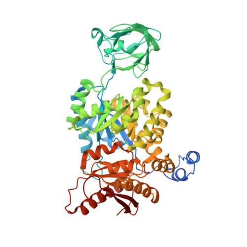
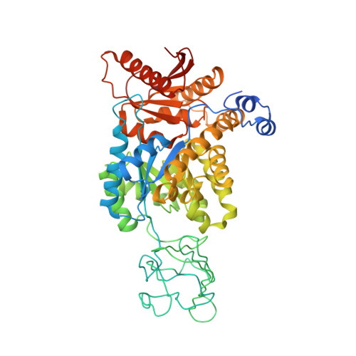
Serine is a natural ligand and allosteric activator of pyruvate kinase M2.

Chaneton, B.Hillmann, P.Zheng, L.Martin, A.C.L.Maddocks, O.D.K.Chokkathukalam, A.Coyle, J.E.Jankevics, A.Holding, F.P.Vousden, K.H.Frezza, C.O'Reilly, M.Gottlieb, E.

(2012) Nature 491: 458-462

  • DOI: https://doi.org/10.1038/nature11540
  • Primary Citation of Related Structures:  
    4B2D

  • PubMed Abstract: 

    Cancer cells exhibit several unique metabolic phenotypes that are critical for cell growth and proliferation. Specifically, they overexpress the M2 isoform of the tightly regulated enzyme pyruvate kinase (PKM2), which controls glycolytic flux, and are highly dependent on de novo biosynthesis of serine and glycine. Here we describe a new rheostat-like mechanistic relationship between PKM2 activity and serine biosynthesis. We show that serine can bind to and activate human PKM2, and that PKM2 activity in cells is reduced in response to serine deprivation. This reduction in PKM2 activity shifts cells to a fuel-efficient mode in which more pyruvate is diverted to the mitochondria and more glucose-derived carbon is channelled into serine biosynthesis to support cell proliferation.


  • Organizational Affiliation

    Cancer Research UK, The Beatson Institute for Cancer Research, Switchback Road, Glasgow, G61 1BD, Scotland, United Kingdom.


Macromolecules
Find similar proteins by:  (by identity cutoff)  |  3D Structure
Entity ID: 1
MoleculeChains Sequence LengthOrganismDetailsImage
PYRUVATE KINASE ISOZYMES M1/M2
A, B, C
548Homo sapiensMutation(s): 0 
EC: 2.7.1.40
UniProt & NIH Common Fund Data Resources
Find proteins for P14618 (Homo sapiens)
Explore P14618 
Go to UniProtKB:  P14618
PHAROS:  P14618
GTEx:  ENSG00000067225 
Entity Groups  
Sequence Clusters30% Identity50% Identity70% Identity90% Identity95% Identity100% Identity
UniProt GroupP14618
Sequence Annotations
Expand
  • Reference Sequence
Find similar proteins by:  (by identity cutoff)  |  3D Structure
Entity ID: 2
MoleculeChains Sequence LengthOrganismDetailsImage
PYRUVATE KINASE ISOZYMES M1/M2548Homo sapiensMutation(s): 0 
EC: 2.7.1.40
UniProt & NIH Common Fund Data Resources
Find proteins for P14618 (Homo sapiens)
Explore P14618 
Go to UniProtKB:  P14618
PHAROS:  P14618
GTEx:  ENSG00000067225 
Entity Groups  
Sequence Clusters30% Identity50% Identity70% Identity90% Identity95% Identity100% Identity
UniProt GroupP14618
Sequence Annotations
Expand
  • Reference Sequence
Small Molecules
Ligands 3 Unique
IDChains Name / Formula / InChI Key2D Diagram3D Interactions
FBP
Query on FBP

Download Ideal Coordinates CCD File 
F [auth A],
I [auth B],
K [auth C],
N [auth D]
1,6-di-O-phosphono-beta-D-fructofuranose
C6 H14 O12 P2
RNBGYGVWRKECFJ-ARQDHWQXSA-N
SER
Query on SER

Download Ideal Coordinates CCD File 
E [auth A],
H [auth B],
J [auth C],
M [auth D]
SERINE
C3 H7 N O3
MTCFGRXMJLQNBG-REOHCLBHSA-N
MG
Query on MG

Download Ideal Coordinates CCD File 
G [auth A],
L [auth C]
MAGNESIUM ION
Mg
JLVVSXFLKOJNIY-UHFFFAOYSA-N
Binding Affinity Annotations 
IDSourceBinding Affinity
SER Binding MOAD:  4B2D Kd: 2.00e+5 (nM) from 1 assay(s)
PDBBind:  4B2D Kd: 2.00e+5 (nM) from 1 assay(s)
Experimental Data & Validation

Experimental Data

  • Method: X-RAY DIFFRACTION
  • Resolution: 2.30 Å
  • R-Value Free: 0.226 
  • R-Value Work: 0.173 
  • R-Value Observed: 0.176 
  • Space Group: P 1 21 1
Unit Cell:
Length ( Å )Angle ( ˚ )
a = 80.611α = 90
b = 151.098β = 102.01
c = 91.791γ = 90
Software Package:
Software NamePurpose
BUSTERrefinement
XDSdata reduction
SCALAdata scaling
CSEARCHphasing

Structure Validation

View Full Validation Report


Ligand Structure Quality Assessment 


Entry History 

Revision History  (Full details and data files)

  • Version 1.0: 2012-10-10
    Type: Initial release
  • Version 1.1: 2012-10-24
    Changes: Database references
  • Version 1.2: 2012-11-21
    Changes: Database references
  • Version 1.3: 2013-02-06
    Changes: Atomic model, Derived calculations, Other
  • Version 1.4: 2018-02-07
    Changes: Database references, Structure summary
  • Version 1.5: 2020-07-29
    Type: Remediation
    Reason: Carbohydrate remediation
    Changes: Data collection, Derived calculations, Other, Structure summary
  • Version 1.6: 2023-12-20
    Changes: Data collection, Database references, Refinement description, Structure summary