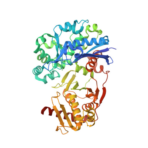
Structure and Function of Human Xylulokinase, an Enzyme with Important Roles in Carbohydrate Metabolism
Bunker, R.D., Bulloch, E.M.M., Dickson, J.M.J., Loomes, K.M., Baker, E.N.(2013) J Biol Chem 288: 1643
- PubMed: 23179721
- DOI: https://doi.org/10.1074/jbc.M112.427997
- Primary Citation of Related Structures:
4BC2, 4BC3, 4BC4, 4BC5 - PubMed Abstract:
D-Xylulokinase (XK; EC 2.7.1.17) catalyzes the ATP-dependent phosphorylation of d-xylulose (Xu) to produce xylulose 5-phosphate (Xu5P). In mammals, XK is the last enzyme in the glucuronate-xylulose pathway, active in the liver and kidneys, and is linked through its product Xu5P to the pentose-phosphate pathway. XK may play an important role in metabolic disease, given that Xu5P is a key regulator of glucose metabolism and lipogenesis. We have expressed the product of a putative human XK gene and identified it as the authentic human d-xylulokinase (hXK). NMR studies with a variety of sugars showed that hXK acts only on d-xylulose, and a coupled photometric assay established its key kinetic parameters as K(m)(Xu) = 24 ± 3 μm and k(cat) = 35 ± 5 s(-1). Crystal structures were determined for hXK, on its own and in complexes with Xu, ADP, and a fluorinated inhibitor. These reveal that hXK has a two-domain fold characteristic of the sugar kinase/hsp70/actin superfamily, with glycerol kinase as its closest relative. Xu binds to domain-I and ADP to domain-II, but in this open form of hXK they are 10 Å apart, implying that a large scale conformational change is required for catalysis. Xu binds in its linear keto-form, sandwiched between a Trp side chain and polar side chains that provide exquisite hydrogen bonding recognition. The hXK structure provides a basis for the design of specific inhibitors with which to probe its roles in sugar metabolism and metabolic disease.
Organizational Affiliation:
Maurice Wilkins Centre for Molecular Biodiscovery and School of Biological Sciences, University of Auckland, Private Bag 92019, Auckland 1142, New Zealand.


