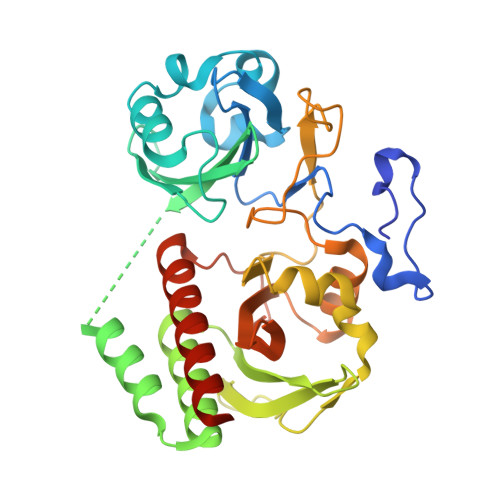
4CQH

Structure of Infrared Fluorescent Protein IFP2.0


Experimental Data Snapshot

  • Method: X-RAY DIFFRACTION
  • Resolution: 1.14 Å
  • R-Value Free: 0.160 
  • R-Value Work: 0.137 
  • R-Value Observed: 0.138 

Starting Model: experimental
View more details

wwPDB Validation   3D Report Full Report


Ligand Structure Quality Assessment 


This is version 1.2 of the entry. See complete history


Literature

An Improved Monomeric Infrared Fluorescent Protein for Neuronal and Tumour Brain Imaging.

Yu, D.Gustafson, W.C.Han, C.Lafaye, C.Noirclerc-Savoye, M.Ge, W.Thayer, D.A.Huang, H.Kornberg, T.B.Royant, A.Jan, L.Y.Jan, Y.N.Weiss, W.A.Shu, X.

(2014) Nat Commun 5: 3626

  • DOI: https://doi.org/10.1038/ncomms4626
  • Primary Citation of Related Structures:  
    4CQH

  • PubMed Abstract: 

    Infrared fluorescent proteins (IFPs) are ideal for in vivo imaging, and monomeric versions of these proteins can be advantageous as protein tags or for sensor development. In contrast to GFP, which requires only molecular oxygen for chromophore maturation, phytochrome-derived IFPs incorporate biliverdin (BV) as the chromophore. However, BV varies in concentration in different cells and organisms. Here we engineered cells to express the haeme oxygenase responsible for BV biosynthesis and a brighter monomeric IFP mutant (IFP2.0). Together, these tools improve the imaging capabilities of IFP2.0 compared with monomeric IFP1.4 and dimeric iRFP. By targeting IFP2.0 to the plasma membrane, we demonstrate robust labelling of neuronal processes in Drosophila larvae. We also show that this strategy improves the sensitivity when imaging brain tumours in whole mice. Our work shows promise in the application of IFPs for protein labelling and in vivo imaging.


  • Organizational Affiliation

    1] Department of Pharmaceutical Chemistry, University of California, San Francisco, California 94158, USA [2] Cardiovascular Research Institute, University of California, San Francisco, California 94158, USA.


Macromolecules
Find similar proteins by:  (by identity cutoff)  |  3D Structure
Entity ID: 1
MoleculeChains Sequence LengthOrganismDetailsImage
BACTERIOPHYTOCHROME329Deinococcus radioduransMutation(s): 16 
EC: 2.7.13.3
UniProt
Find proteins for Q9RZA4 (Deinococcus radiodurans (strain ATCC 13939 / DSM 20539 / JCM 16871 / CCUG 27074 / LMG 4051 / NBRC 15346 / NCIMB 9279 / VKM B-1422 / R1))
Explore Q9RZA4 
Go to UniProtKB:  Q9RZA4
Entity Groups  
Sequence Clusters30% Identity50% Identity70% Identity90% Identity95% Identity100% Identity
UniProt GroupQ9RZA4
Sequence Annotations
Expand
  • Reference Sequence
Small Molecules
Ligands 2 Unique
IDChains Name / Formula / InChI Key2D Diagram3D Interactions
LBV
Query on LBV

Download Ideal Coordinates CCD File 
B [auth A]3-[2-[(Z)-[3-(2-carboxyethyl)-5-[(Z)-(4-ethenyl-3-methyl-5-oxidanylidene-pyrrol-2-ylidene)methyl]-4-methyl-pyrrol-1-ium -2-ylidene]methyl]-5-[(Z)-[(3E)-3-ethylidene-4-methyl-5-oxidanylidene-pyrrolidin-2-ylidene]methyl]-4-methyl-1H-pyrrol-3- yl]propanoic acid
C33 H37 N4 O6
DKMLMZVDTGOEGU-ISEYCTJISA-O
NA
Query on NA

Download Ideal Coordinates CCD File 
C [auth A]SODIUM ION
Na
FKNQFGJONOIPTF-UHFFFAOYSA-N
Experimental Data & Validation

Experimental Data

  • Method: X-RAY DIFFRACTION
  • Resolution: 1.14 Å
  • R-Value Free: 0.160 
  • R-Value Work: 0.137 
  • R-Value Observed: 0.138 
  • Space Group: C 1 2 1
Unit Cell:
Length ( Å )Angle ( ˚ )
a = 95.692α = 90
b = 52.912β = 91.37
c = 66.316γ = 90
Software Package:
Software NamePurpose
REFMACrefinement
XDSdata reduction
Aimlessdata scaling
PHASERphasing

Structure Validation

View Full Validation Report


Ligand Structure Quality Assessment 


Entry History 

Deposition Data

Revision History  (Full details and data files)

  • Version 1.0: 2014-05-28
    Type: Initial release
  • Version 1.1: 2023-12-20
    Changes: Data collection, Database references, Derived calculations, Other, Refinement description, Structure summary
  • Version 1.2: 2024-11-06
    Changes: Structure summary