4CU4

FhuA from E. coli in complex with the lasso peptide microcin J25 (MccJ25)


Experimental Data Snapshot

  • Method: X-RAY DIFFRACTION
  • Resolution: 2.30 Å
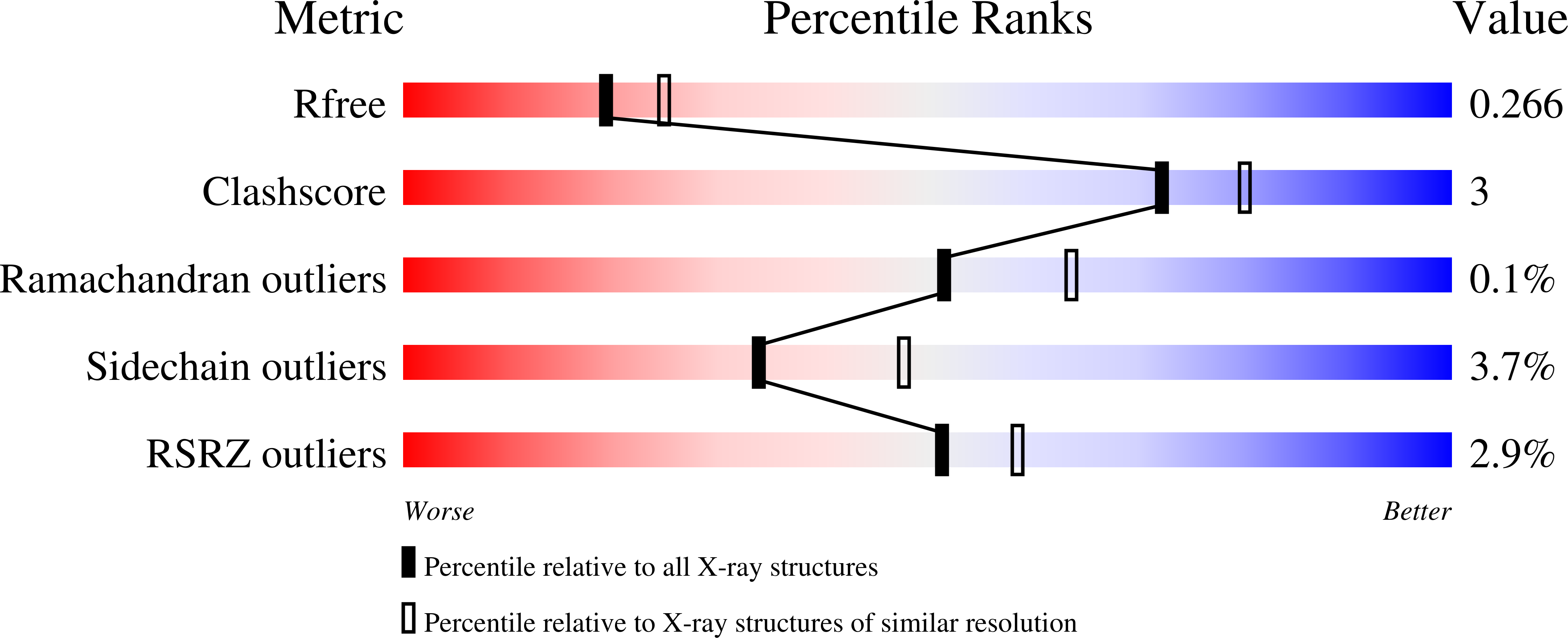
  • R-Value Free: 0.255 
  • R-Value Work: 0.212 
  • R-Value Observed: 0.214 

Starting Model: experimental
View more details

wwPDB Validation   3D Report Full Report


Ligand Structure Quality Assessment 


This is version 2.2 of the entry. See complete history


Literature

Structural Basis for Hijacking Siderophore Receptors by Antimicrobial Lasso Peptides.

Mathavan, I.Zirah, S.Mehmood, S.Choudhury, H.G.Goulard, C.Li, Y.Robinson, C.V.Rebuffat, S.Beis, K.

(2014) Nat Chem Biol 10: 340

  • DOI: https://doi.org/10.1038/nchembio.1499
  • Primary Citation of Related Structures:  
    4CU4

  • PubMed Abstract: 

    The lasso peptide microcin J25 is known to hijack the siderophore receptor FhuA for initiating internalization. Here, we provide what is to our knowledge the first structural evidence on the recognition mechanism, and our biochemical data show that another closely related lasso peptide cannot interact with FhuA. Our work provides an explanation on the narrow activity spectrum of lasso peptides and opens the path to the development of new antibacterials.


  • Organizational Affiliation

    1] Department of Life Sciences, Imperial College London, London, UK. [2] Membrane Protein Lab, Diamond Light Source, Harwell Science and Innovation Campus, Chilton, Oxfordshire, UK. [3] Rutherford Appleton Laboratory, Research Complex at Harwell, Didcot, Oxfordshire, UK.


Macromolecules
Find similar proteins by:  (by identity cutoff)  |  3D Structure
Entity ID: 1
MoleculeChains Sequence LengthOrganismDetailsImage
FERRICHROME-IRON RECEPTOR706Escherichia coli str. K-12 substr. MG1655Mutation(s): 0 
Membrane Entity: Yes 
UniProt
Find proteins for P06971 (Escherichia coli (strain K12))
Explore P06971 
Go to UniProtKB:  P06971
Entity Groups  
Sequence Clusters30% Identity50% Identity70% Identity90% Identity95% Identity100% Identity
UniProt GroupP06971
Sequence Annotations
Expand
  • Reference Sequence

Find similar proteins by:  Sequence   |   3D Structure  

Entity ID: 2
MoleculeChains Sequence LengthOrganismDetailsImage
MICROCIN J2521Escherichia coli str. K-12 substr. MC4100Mutation(s): 0 
UniProt
Find proteins for Q9X2V7 (Escherichia coli)
Explore Q9X2V7 
Go to UniProtKB:  Q9X2V7
Entity Groups  
Sequence Clusters30% Identity50% Identity70% Identity90% Identity95% Identity100% Identity
UniProt GroupQ9X2V7
Sequence Annotations
Expand
  • Reference Sequence
Oligosaccharides

Help

Entity ID: 3
MoleculeChains Length2D Diagram Glycosylation3D Interactions
L-glycero-alpha-D-manno-heptopyranose-(1-3)-L-glycero-alpha-D-manno-heptopyranose-(1-5)-[3-deoxy-alpha-D-manno-oct-2-ulopyranosonic acid-(2-4)]3-deoxy-alpha-D-manno-oct-2-ulopyranosonic acid-(2-6)-2-amino-2-deoxy-beta-D-glucopyranose-(1-6)-2-amino-2-deoxy-alpha-D-glucopyranose
C
6N/A
Glycosylation Resources
GlyTouCan:  G23424DU
GlyCosmos:  G23424DU
Small Molecules
Ligands 7 Unique
IDChains Name / Formula / InChI Key2D Diagram3D Interactions
3PH
Query on 3PH

Download Ideal Coordinates CCD File 
BA [auth A]1,2-DIACYL-GLYCEROL-3-SN-PHOSPHATE
C39 H77 O8 P
YFWHNAWEOZTIPI-DIPNUNPCSA-N
FTT
Query on FTT

Download Ideal Coordinates CCD File 
D [auth A],
E [auth A],
F [auth A],
H [auth A]
3-HYDROXY-TETRADECANOIC ACID
C14 H28 O3
ATRNZOYKSNPPBF-CYBMUJFWSA-N
LDA
Query on LDA

Download Ideal Coordinates CCD File 
AA [auth A]
CA [auth A]
DA [auth A]
J [auth A]
K [auth A]
AA [auth A],
CA [auth A],
DA [auth A],
J [auth A],
K [auth A],
L [auth A],
M [auth A],
N [auth A],
O [auth A],
P [auth A],
Q [auth A],
R [auth A],
S [auth A],
T [auth A],
U [auth A],
V [auth A],
W [auth A],
X [auth A],
Y [auth A],
Z [auth A]
LAURYL DIMETHYLAMINE-N-OXIDE
C14 H31 N O
SYELZBGXAIXKHU-UHFFFAOYSA-N
MYR
Query on MYR

Download Ideal Coordinates CCD File 
I [auth A]MYRISTIC ACID
C14 H28 O2
TUNFSRHWOTWDNC-UHFFFAOYSA-N
DAO
Query on DAO

Download Ideal Coordinates CCD File 
G [auth A]LAURIC ACID
C12 H24 O2
POULHZVOKOAJMA-UHFFFAOYSA-N
DPO
Query on DPO

Download Ideal Coordinates CCD File 
EA [auth A],
GA [auth A]
DIPHOSPHATE
O7 P2
XPPKVPWEQAFLFU-UHFFFAOYSA-J
PO4
Query on PO4

Download Ideal Coordinates CCD File 
FA [auth A],
HA [auth A]
PHOSPHATE ION
O4 P
NBIIXXVUZAFLBC-UHFFFAOYSA-K
Biologically Interesting Molecules (External Reference) 1 Unique
Experimental Data & Validation

Experimental Data

  • Method: X-RAY DIFFRACTION
  • Resolution: 2.30 Å
  • R-Value Free: 0.255 
  • R-Value Work: 0.212 
  • R-Value Observed: 0.214 
  • Space Group: C 2 2 21
Unit Cell:
Length ( Å )Angle ( ˚ )
a = 95.98α = 90
b = 277.05β = 90
c = 106.76γ = 90
Software Package:
Software NamePurpose
BUSTERrefinement
xia2data reduction
SCALAdata scaling
PHASERphasing

Structure Validation

View Full Validation Report


Ligand Structure Quality Assessment 


Entry History 

Deposition Data

Revision History  (Full details and data files)

  • Version 1.0: 2014-04-09
    Type: Initial release
  • Version 1.1: 2014-04-16
    Changes: Database references
  • Version 1.2: 2014-04-30
    Changes: Database references
  • Version 1.3: 2018-06-13
    Changes: Data collection, Structure summary
  • Version 1.4: 2019-07-31
    Changes: Advisory, Data collection, Derived calculations
  • Version 1.5: 2020-07-01
    Changes: Data collection, Other
  • Version 2.0: 2020-07-29
    Type: Remediation
    Reason: Carbohydrate remediation
    Changes: Advisory, Atomic model, Data collection, Derived calculations, Structure summary
  • Version 2.1: 2023-12-20
    Changes: Data collection, Database references, Derived calculations, Refinement description, Structure summary
  • Version 2.2: 2024-11-13
    Changes: Structure summary