4D1X | pdb_00004d1x

CDK2 in complex with Luciferin


Experimental Data Snapshot

  • Method: X-RAY DIFFRACTION
  • Resolution: 2.10 Å
  • R-Value Free: 
    0.238 (Depositor), 0.239 (DCC) 
  • R-Value Work: 
    0.194 (Depositor), 0.195 (DCC) 
  • R-Value Observed: 
    0.196 (Depositor) 

Starting Model: experimental
View more details

wwPDB Validation 3D Report Full Report

Validation slider image for 4D1X

Ligand Structure Quality Assessment 


This is version 1.2 of the entry. See complete history

Literature

Luciferin and Derivatives as a Dyrk Selective Scaffold for the Design of Protein Kinase Inhibitors.

Rothweiler, U.Eriksson, J.Stensen, W.Leeson, F.Engh, R.A.Svendsen, J.S.

(2015) Eur J Med Chem 94: 140

  • DOI: https://doi.org/10.1016/j.ejmech.2015.02.035
  • Primary Citation Related Structures: 
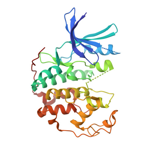
    4D1X, 4D1Z

  • PubMed Abstract: 

    D-Luciferin is widely used as a substrate in luciferase catalysed bioluminescence assays for in vitro studies. However, little is known about cross reactivity and potential interference of D-luciferin with other enzymes. We serendipitously found that firefly luciferin inhibited the CDK2/Cyclin A protein kinase. Inhibition profiling of D-luciferin over a 103-protein kinase panel showed significant inhibition of a small set of protein kinases, in particular the DYRK-family, but also other members of the CMGC-group, including ERK8 and CK2. Inhibition profiling on a 16-member focused library derived from D-luciferin confirms that D-luciferin represents a DYRK-selective chemotype of fragment-like molecular weight. Thus, observation of its inhibitory activity and the initial SAR information reported here promise to be useful for further design of protein kinase inhibitors with related scaffolds.


  • Organizational Affiliation
    • The Norwegian Structural Biology Centre, Department of Chemistry, UiT The Arctic University of Norway, N-9037 Tromsø, Norway.

Macromolecule Content 

  • Total Structure Weight: 34.26 kDa 
  • Atom Count: 2,298 
  • Modeled Residue Count: 274 
  • Deposited Residue Count: 298 
  • Unique protein chains: 1

Macromolecules

Find similar proteins by:|  3D Structure
Entity ID: 1
MoleculeChains  Sequence LengthOrganismDetailsImage
CYCLIN-DEPENDENT KINASE 2298Homo sapiensMutation(s): 0 
EC: 2.7.11.22
UniProt & NIH Common Fund Data Resources
Find proteins for P24941 (Homo sapiens)
Explore P24941 
Go to UniProtKB:  P24941
PHAROS:  P24941
GTEx:  ENSG00000123374 
Entity Groups
Sequence Clusters30% Identity50% Identity70% Identity90% Identity95% Identity100% Identity
UniProt GroupP24941
Sequence Annotations
Expand
Reference Sequence

Small Molecules

Ligands 1 Unique
IDChains Name / Formula / InChI Key2D Diagram3D Interactions
ESJ

Query on ESJ


Download:Ideal Coordinates CCD File
B [auth A](4S)-2-(6-hydroxy-1,3-benzothiazol-2-yl)-4,5-dihydro-1,3-thiazole-4-carboxylic acid
C11 H8 N2 O3 S2
BJGNCJDXODQBOB-SSDOTTSWSA-N

Experimental Data & Validation

Experimental Data

  • Method: X-RAY DIFFRACTION
  • Resolution: 2.10 Å
  • R-Value Free:  0.238 (Depositor), 0.239 (DCC) 
  • R-Value Work:  0.194 (Depositor), 0.195 (DCC) 
  • R-Value Observed: 0.196 (Depositor) 
Space Group: P 21 21 21
Unit Cell:
Length ( Å )Angle ( ˚ )
a = 53.31α = 90
b = 72.12β = 90
c = 72.29γ = 90
Software Package:
Software NamePurpose
PHENIXrefinement
XDSdata reduction
XDSdata scaling
MOLREPphasing

Structure Validation

View Full Validation Report


Ligand Structure Quality Assessment 


Entry History 

Deposition Data

Revision History  (Full details and data files)

  • Version 1.0: 2015-03-18
    Type: Initial release
  • Version 1.1: 2015-03-25
    Changes: Database references
  • Version 1.2: 2023-12-20
    Changes: Data collection, Database references, Derived calculations, Other, Refinement description