4D2I

Crystal structure of the HerA hexameric DNA translocase from Sulfolobus solfataricus bound to AMP-PNP


Experimental Data Snapshot

  • Method: X-RAY DIFFRACTION
  • Resolution: 2.84 Å
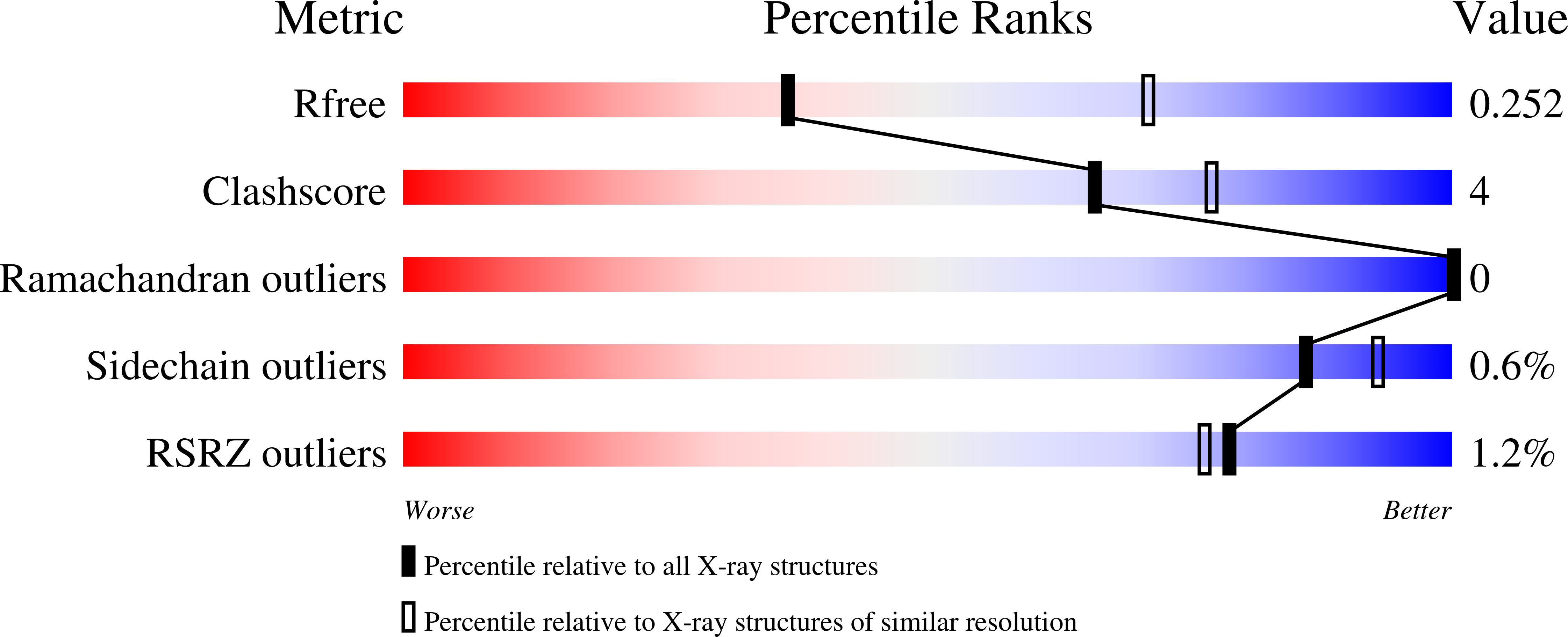
  • R-Value Free: 
    0.254 (Depositor), 0.250 (DCC) 
  • R-Value Work: 
    0.211 (Depositor), 0.220 (DCC) 
  • R-Value Observed: 
    0.213 (Depositor) 

wwPDB Validation   3D Report Full Report


Ligand Structure Quality Assessment 

Created with Raphaël 2.3.0Worse 01 BetterLigand structure goodness of fit to experimental dataBest fitted ANPClick on this verticalbar to view details

This is version 1.1 of the entry. See complete history


Literature

Structure of the Hexameric Hera ATPase Reveals a Mechanism of Translocation-Coupled DNA-End Processing in Archaea

Rzechorzek, N.J.Blackwood, J.K.Bray, S.M.Maman, J.D.Pellegrini, L.Robinson, N.P.

(2014) Nat Commun 5: 5506

  • DOI: https://doi.org/10.1038/ncomms6506
  • Primary Citation of Related Structures:  
    4D2I

  • PubMed Abstract: 

    The HerA ATPase cooperates with the NurA nuclease and the Mre11-Rad50 complex for the repair of double-strand DNA breaks in thermophilic archaea. Here we extend our structural knowledge of this minimal end-resection apparatus by presenting the first crystal structure of hexameric HerA. The full-length structure visualizes at atomic resolution the N-terminal HerA-ATP synthase domain and a conserved C-terminal extension, which acts as a physical brace between adjacent protomers. The brace also interacts in trans with nucleotide-binding residues of the neighbouring subunit. Our observations support a model in which the coaxial interaction of the HerA ring with the toroidal NurA dimer generates a continuous channel traversing the complex. HerA-driven translocation would propel the DNA towards the narrow annulus of NurA, leading to duplex melting and nucleolytic digestion. This system differs substantially from the bacterial end-resection paradigms. Our findings suggest a novel mode of DNA-end processing by this integrated archaeal helicase-nuclease machine.


  • Organizational Affiliation

    Department of Biochemistry, University of Cambridge, Tennis Court Road, Cambridge CB2 1GA, UK.


Macromolecules
Find similar proteins by:  (by identity cutoff)  |  3D Structure
Entity ID: 1
MoleculeChains Sequence LengthOrganismDetailsImage
HERA
A, B
500Saccharolobus solfataricus P2Mutation(s): 10 
EC: 3.6.4.12
UniProt
Find proteins for Q97WG8 (Saccharolobus solfataricus (strain ATCC 35092 / DSM 1617 / JCM 11322 / P2))
Explore Q97WG8 
Go to UniProtKB:  Q97WG8
Entity Groups  
Sequence Clusters30% Identity50% Identity70% Identity90% Identity95% Identity100% Identity
UniProt GroupQ97WG8
Sequence Annotations
Expand
  • Reference Sequence
Experimental Data & Validation

Experimental Data

  • Method: X-RAY DIFFRACTION
  • Resolution: 2.84 Å
  • R-Value Free:  0.254 (Depositor), 0.250 (DCC) 
  • R-Value Work:  0.211 (Depositor), 0.220 (DCC) 
  • R-Value Observed: 0.213 (Depositor) 
Space Group: H 3
Unit Cell:
Length ( Å )Angle ( ˚ )
a = 150.21α = 90
b = 150.21β = 90
c = 140.54γ = 120
Software Package:
Software NamePurpose
PHENIXrefinement
XDSdata reduction
XDSdata scaling
PHASERphasing

Structure Validation

View Full Validation Report


Ligand Structure Quality Assessment 

Created with Raphaël 2.3.0Worse 01 BetterLigand structure goodness of fit to experimental dataBest fitted ANPClick on this verticalbar to view details

Entry History 

Deposition Data

Revision History  (Full details and data files)

  • Version 1.0: 2014-12-03
    Type: Initial release
  • Version 1.1: 2024-05-08
    Changes: Data collection, Database references, Derived calculations, Other