4D5E

Crystal Structure of recombinant wildtype CDH


Experimental Data Snapshot

  • Method: X-RAY DIFFRACTION
  • Resolution: 1.43 Å
  • R-Value Free: 
    0.147 (Depositor), 0.160 (DCC) 
  • R-Value Work: 
    0.122 (Depositor), 0.140 (DCC) 
  • R-Value Observed: 
    0.123 (Depositor) 

wwPDB Validation   3D Report Full Report


Ligand Structure Quality Assessment 


This is version 1.3 of the entry. See complete history


Literature

Extended Reaction Scope of Thiamine Diphosphate Dependent Cyclohexane-1,2-Dione Hydrolase: From C-C Bond Cleavage to C-C Bond Ligation.

Loschonsky, S.Wacker, T.Waltzer, S.Giovannini, P.P.Mcleish, M.J.Andrade, S.L.A.Muller, M.

(2014) Angew Chem Int Ed Engl 53: 14402

  • DOI: https://doi.org/10.1002/anie.201408287
  • Primary Citation of Related Structures:  
    4D5E, 4D5G

  • PubMed Abstract: 

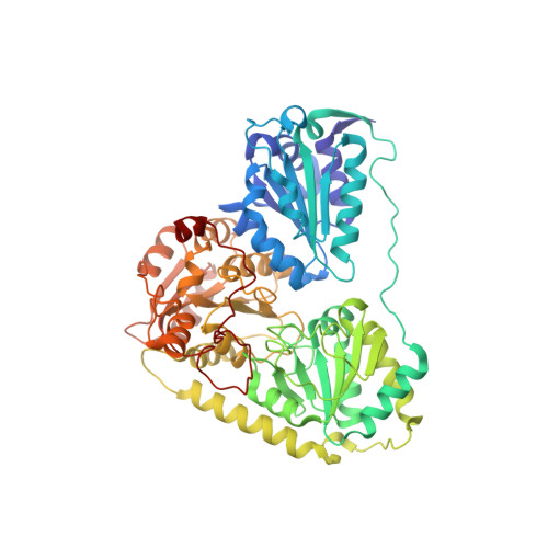
    ThDP-dependent cyclohexane-1,2-dione hydrolase (CDH) catalyzes the CC bond cleavage of cyclohexane-1,2-dione to 6-oxohexanoate, and the asymmetric benzoin condensation between benzaldehyde and pyruvate. One of the two reactivities of CDH was selectively knocked down by mutation experiments. CDH-H28A is much less able to catalyze the CC bond formation, while the ability for CC bond cleavage is still intact. The double variant CDH-H28A/N484A shows the opposite behavior and catalyzes the addition of pyruvate to cyclohexane-1,2-dione, resulting in the formation of a tertiary alcohol. Several acyloins of tertiary alcohols are formed with 54-94 % enantiomeric excess. In addition to pyruvate, methyl pyruvate and butane-2,3-dione are alternative donor substrates for CC bond formation. Thus, the very rare aldehyde-ketone cross-benzoin reaction has been solved by design of an enzyme variant.


  • Organizational Affiliation

    Institute of Pharmaceutical Sciences, Albert-Ludwigs-Universität Freiburg, Albertstrasse 25, 79104 Freiburg (Germany).


Macromolecules
Find similar proteins by:  (by identity cutoff)  |  3D Structure
Entity ID: 1
MoleculeChains Sequence LengthOrganismDetailsImage
CYCLOHEXANE-1,2-DIONE HYDROLASE
A, B
589Azoarcus oleariusMutation(s): 0 
EC: 3.7.1.11
UniProt
Find proteins for P0CH62 (Azoarcus sp)
Explore P0CH62 
Go to UniProtKB:  P0CH62
Entity Groups  
Sequence Clusters30% Identity50% Identity70% Identity90% Identity95% Identity100% Identity
UniProt GroupP0CH62
Sequence Annotations
Expand
  • Reference Sequence
Small Molecules
Ligands 10 Unique
IDChains Name / Formula / InChI Key2D Diagram3D Interactions
FAD
Query on FAD

Download Ideal Coordinates CCD File 
C [auth A],
P [auth B]
FLAVIN-ADENINE DINUCLEOTIDE
C27 H33 N9 O15 P2
VWWQXMAJTJZDQX-UYBVJOGSSA-N
TPP
Query on TPP

Download Ideal Coordinates CCD File 
D [auth A],
Q [auth B]
THIAMINE DIPHOSPHATE
C12 H19 N4 O7 P2 S
AYEKOFBPNLCAJY-UHFFFAOYSA-O
2PE
Query on 2PE

Download Ideal Coordinates CCD File 
M [auth A],
V [auth B],
X [auth B]
NONAETHYLENE GLYCOL
C18 H38 O10
YZUUTMGDONTGTN-UHFFFAOYSA-N
P6G
Query on P6G

Download Ideal Coordinates CCD File 
H [auth A],
J [auth A]
HEXAETHYLENE GLYCOL
C12 H26 O7
IIRDTKBZINWQAW-UHFFFAOYSA-N
1PG
Query on 1PG

Download Ideal Coordinates CCD File 
I [auth A]2-(2-{2-[2-(2-METHOXY-ETHOXY)-ETHOXY]-ETHOXY}-ETHOXY)-ETHANOL
C11 H24 O6
SLNYBUIEAMRFSZ-UHFFFAOYSA-N
MES
Query on MES

Download Ideal Coordinates CCD File 
E [auth A],
R [auth B]
2-(N-MORPHOLINO)-ETHANESULFONIC ACID
C6 H13 N O4 S
SXGZJKUKBWWHRA-UHFFFAOYSA-N
PG4
Query on PG4

Download Ideal Coordinates CCD File 
AA [auth B]
BA [auth B]
CA [auth B]
K [auth A]
N [auth A]
AA [auth B],
BA [auth B],
CA [auth B],
K [auth A],
N [auth A],
O [auth A],
W [auth B],
Y [auth B],
Z [auth B]
TETRAETHYLENE GLYCOL
C8 H18 O5
UWHCKJMYHZGTIT-UHFFFAOYSA-N
PEG
Query on PEG

Download Ideal Coordinates CCD File 
L [auth A]DI(HYDROXYETHYL)ETHER
C4 H10 O3
MTHSVFCYNBDYFN-UHFFFAOYSA-N
CL
Query on CL

Download Ideal Coordinates CCD File 
F [auth A],
S [auth B]
CHLORIDE ION
Cl
VEXZGXHMUGYJMC-UHFFFAOYSA-M
MG
Query on MG

Download Ideal Coordinates CCD File 
G [auth A],
T [auth B],
U [auth B]
MAGNESIUM ION
Mg
JLVVSXFLKOJNIY-UHFFFAOYSA-N
Experimental Data & Validation

Experimental Data

  • Method: X-RAY DIFFRACTION
  • Resolution: 1.43 Å
  • R-Value Free:  0.147 (Depositor), 0.160 (DCC) 
  • R-Value Work:  0.122 (Depositor), 0.140 (DCC) 
  • R-Value Observed: 0.123 (Depositor) 
Space Group: P 41 21 2
Unit Cell:
Length ( Å )Angle ( ˚ )
a = 123.188α = 90
b = 123.188β = 90
c = 143.07γ = 90
Software Package:
Software NamePurpose
REFMACrefinement
XDSdata reduction
SCALAdata scaling

Structure Validation

View Full Validation Report


Ligand Structure Quality Assessment 


Entry History 

Deposition Data

Revision History  (Full details and data files)

  • Version 1.0: 2015-01-21
    Type: Initial release
  • Version 1.1: 2015-05-06
    Changes: Database references
  • Version 1.2: 2017-08-23
    Changes: Advisory, Data collection
  • Version 1.3: 2024-05-08
    Changes: Advisory, Data collection, Database references, Derived calculations, Other