4FC2 | pdb_00004fc2

Crystal structure of mouse poly(ADP-ribose) glycohydrolase (PARG) catalytic domain


Experimental Data Snapshot

  • Method: X-RAY DIFFRACTION
  • Resolution: 1.91 Å
  • R-Value Free: 
    0.214 (Depositor), 0.223 (DCC) 
  • R-Value Work: 
    0.174 (Depositor), 0.182 (DCC) 
  • R-Value Observed: 
    0.176 (Depositor) 

wwPDB Validation 3D Report Full Report

Validation slider image for 4FC2

This is version 1.4 of the entry. See complete history

Literature

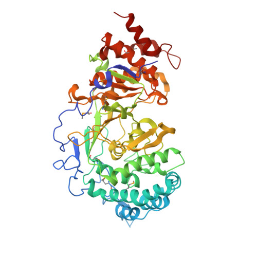
Crystallographic and biochemical analysis of the mouse poly(ADP-ribose) glycohydrolase.

Wang, Z.Gagne, J.P.Poirier, G.G.Xu, W.

(2014) PLoS One 9: e86010-e86010

  • DOI: https://doi.org/10.1371/journal.pone.0086010
  • Primary Citation Related Structures: 
    4FC2, 4N9Y, 4N9Z, 4NA0, 4NA4, 4NA5, 4NA6

  • PubMed Abstract: 

    Protein poly(ADP-ribosyl)ation (PARylation) regulates a number of important cellular processes. Poly(ADP-ribose) glycohydrolase (PARG) is the primary enzyme responsible for hydrolyzing the poly(ADP-ribose) (PAR) polymer in vivo. Here we report crystal structures of the mouse PARG (mPARG) catalytic domain, its complexes with ADP-ribose (ADPr) and a PARG inhibitor ADP-HPD, as well as four PARG catalytic residues mutants. With these structures and biochemical analysis of 20 mPARG mutants, we provide a structural basis for understanding how the PAR polymer is recognized and hydrolyzed by mPARG. The structures and activity complementation experiment also suggest how the N-terminal flexible peptide preceding the PARG catalytic domain may regulate the enzymatic activity of PARG. This study contributes to our understanding of PARG catalytic and regulatory mechanisms as well as the rational design of PARG inhibitors.


  • Organizational Affiliation
    • Department of Biological Structure, University of Washington, Seattle, Washington, United States of America.

Macromolecule Content 

  • Total Structure Weight: 244.74 kDa 
  • Atom Count: 18,082 
  • Modeled Residue Count: 2,031 
  • Deposited Residue Count: 2,084 
  • Unique protein chains: 1

Macromolecules

Find similar proteins by:|  3D Structure
Entity ID: 1
MoleculeChains  Sequence LengthOrganismDetailsImage
Poly(ADP-ribose) glycohydrolase
A, B, C, D
521Mus musculusMutation(s): 0 
Gene Names: Parg
EC: 3.2.1.143
UniProt & NIH Common Fund Data Resources
Find proteins for O88622 (Mus musculus)
Explore O88622 
Go to UniProtKB:  O88622
IMPC:  MGI:1347094
Entity Groups
Sequence Clusters30% Identity50% Identity70% Identity90% Identity95% Identity100% Identity
UniProt GroupO88622
Sequence Annotations
Expand
Reference Sequence

Small Molecules

Ligands 1 Unique
IDChains Name / Formula / InChI Key2D Diagram3D Interactions
SO4

Query on SO4


Download:Ideal Coordinates CCD File
AA [auth D]
BA [auth D]
CA [auth D]
DA [auth D]
E [auth A]
AA [auth D],
BA [auth D],
CA [auth D],
DA [auth D],
E [auth A],
EA [auth D],
F [auth A],
FA [auth D],
G [auth A],
H [auth A],
I [auth A],
J [auth A],
K [auth A],
L [auth B],
M [auth B],
N [auth B],
O [auth B],
P [auth B],
Q [auth B],
R [auth B],
S [auth C],
T [auth C],
U [auth C],
V [auth C],
W [auth C],
X [auth C],
Y [auth C],
Z [auth D]
SULFATE ION
O4 S
QAOWNCQODCNURD-UHFFFAOYSA-L
Modified Residues  1 Unique
IDChains TypeFormula2D DiagramParent
MSE
Query on MSE
A, B, C, D
L-PEPTIDE LINKINGC5 H11 N O2 SeMET

Experimental Data & Validation

Experimental Data

  • Method: X-RAY DIFFRACTION
  • Resolution: 1.91 Å
  • R-Value Free:  0.214 (Depositor), 0.223 (DCC) 
  • R-Value Work:  0.174 (Depositor), 0.182 (DCC) 
  • R-Value Observed: 0.176 (Depositor) 
Space Group: P 1
Unit Cell:
Length ( Å )Angle ( ˚ )
a = 67.141α = 81.64
b = 90.401β = 88.41
c = 104.696γ = 89.36
Software Package:
Software NamePurpose
REFMACrefinement
PDB_EXTRACTdata extraction
ADSCdata collection
HKL-2000data reduction
HKL-2000data scaling
SOLVEphasing

Structure Validation

View Full Validation Report


Entry History 

Deposition Data

Revision History  (Full details and data files)

  • Version 1.0: 2012-06-06
    Type: Initial release
  • Version 1.1: 2014-01-29
    Changes: Database references
  • Version 1.2: 2018-10-10
    Changes: Data collection, Database references, Structure summary
  • Version 1.3: 2018-10-17
    Changes: Data collection, Database references, Structure summary
  • Version 1.4: 2024-11-27
    Changes: Data collection, Database references, Derived calculations, Structure summary