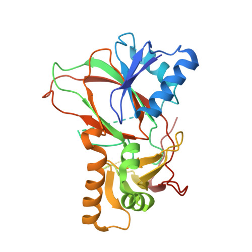
4FFA

Sulfatase from Mycobacterium tuberculosis


Experimental Data Snapshot

  • Method: X-RAY DIFFRACTION
  • Resolution: 2.50 Å
  • R-Value Free: 0.272 
  • R-Value Work: 0.217 
  • R-Value Observed: 0.220 

Starting Model: experimental
View more details

wwPDB Validation   3D Report Full Report


This is version 1.3 of the entry. See complete history


Literature

Mycobacterium tuberculosis Rv3406 Is a Type II Alkyl Sulfatase Capable of Sulfate Scavenging.

Sogi, K.M.Gartner, Z.J.Breidenbach, M.A.Appel, M.J.Schelle, M.W.Bertozzi, C.R.

(2013) PLoS One 8: e65080-e65080

  • DOI: https://doi.org/10.1371/journal.pone.0065080
  • Primary Citation of Related Structures:  
    4FFA

  • PubMed Abstract: 

    The genome of Mycobacterium tuberculosis (Mtb) encodes nine putative sulfatases, none of which have a known function or substrate. Here, we characterize Mtb's single putative type II sulfatase, Rv3406, as a non-heme iron (II) and α-ketoglutarate-dependent dioxygenase that catalyzes the oxidation and subsequent cleavage of alkyl sulfate esters. Rv3406 was identified based on its homology to the alkyl sulfatase AtsK from Pseudomonas putida. Using an in vitro biochemical assay, we confirmed that Rv3406 is a sulfatase with a preference for alkyl sulfate substrates similar to those processed by AtsK. We determined the crystal structure of the apo Rv3406 sulfatase at 2.5 Å. The active site residues of Rv3406 and AtsK are essentially superimposable, suggesting that the two sulfatases share the same catalytic mechanism. Finally, we generated an Rv3406 mutant (Δrv3406) in Mtb to study the sulfatase's role in sulfate scavenging. The Δrv3406 strain did not replicate in minimal media with 2-ethyl hexyl sulfate as the sole sulfur source, in contrast to wild type Mtb or the complemented strain. We conclude that Rv3406 is an iron and α-ketoglutarate-dependent sulfate ester dioxygenase that has unique substrate specificity that is likely distinct from other Mtb sulfatases.


  • Organizational Affiliation

    Department of Chemistry, University of California, Berkeley, California, United States of America.


Macromolecules
Find similar proteins by:  (by identity cutoff)  |  3D Structure
Entity ID: 1
MoleculeChains Sequence LengthOrganismDetailsImage
Rv3406 alkyl sulfatase
A, B, C, D
298Mycobacterium tuberculosisMutation(s): 0 
Gene Names: MT3514MTCY78.22cRv3406
EC: 1.14.11.77
UniProt
Find proteins for P9WKZ1 (Mycobacterium tuberculosis (strain ATCC 25618 / H37Rv))
Explore P9WKZ1 
Go to UniProtKB:  P9WKZ1
Entity Groups  
Sequence Clusters30% Identity50% Identity70% Identity90% Identity95% Identity100% Identity
UniProt GroupP9WKZ1
Sequence Annotations
Expand
  • Reference Sequence
Small Molecules
Ligands 1 Unique
IDChains Name / Formula / InChI Key2D Diagram3D Interactions
NO3
Query on NO3

Download Ideal Coordinates CCD File 
E [auth A]NITRATE ION
N O3
NHNBFGGVMKEFGY-UHFFFAOYSA-N
Experimental Data & Validation

Experimental Data

  • Method: X-RAY DIFFRACTION
  • Resolution: 2.50 Å
  • R-Value Free: 0.272 
  • R-Value Work: 0.217 
  • R-Value Observed: 0.220 
  • Space Group: P 21 21 21
Unit Cell:
Length ( Å )Angle ( ˚ )
a = 64.752α = 90
b = 128.472β = 90
c = 139.332γ = 90
Software Package:
Software NamePurpose
ADSCdata collection
PHASERphasing
PHENIXrefinement
HKL-2000data reduction
SCALEPACKdata scaling

Structure Validation

View Full Validation Report


Entry History 

Deposition Data

Revision History  (Full details and data files)

  • Version 1.0: 2013-06-05
    Type: Initial release
  • Version 1.1: 2013-06-26
    Changes: Database references
  • Version 1.2: 2023-09-13
    Changes: Data collection, Database references, Derived calculations, Refinement description
  • Version 1.3: 2024-11-06
    Changes: Structure summary