4G0C | pdb_00004g0c

Neutron structure of acetazolamide-bound human carbonic anhydrase II reveal molecular details of drug binding.


Experimental Data Snapshot

  • Method: NEUTRON DIFFRACTION
  • Resolution: 2.00 Å
  • R-Value Free: 
    0.283 (Depositor) 
  • R-Value Work: 
    0.267 (Depositor) 

Starting Model: experimental
View more details

wwPDB Validation 3D Report Full Report

Validation slider image for 4G0C

This is version 1.4 of the entry. See complete history

Literature

Neutron diffraction of acetazolamide-bound human carbonic anhydrase II reveals atomic details of drug binding.

Fisher, S.Z.Aggarwal, M.Kovalevsky, A.Y.Silverman, D.N.McKenna, R.

(2012) J Am Chem Soc 134: 14726-14729

  • DOI: https://doi.org/10.1021/ja3068098
  • Primary Citation Related Structures: 
    4G0C

  • PubMed Abstract: 

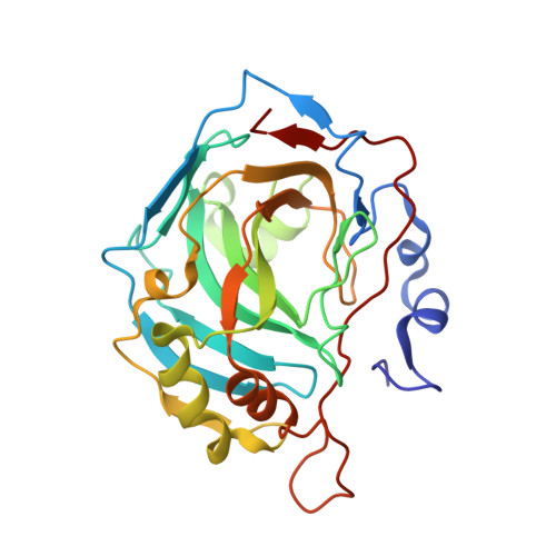
    Carbonic anhydrases (CAs) catalyze the hydration of CO(2) forming HCO(3)(-) and a proton, an important reaction for many physiological processes including respiration, fluid secretion, and pH regulation. As such, CA isoforms are prominent clinical targets for treating various diseases. The clinically used acetazolamide (AZM) is a sulfonamide that binds with high affinity to human CA isoform II (HCA II). There are several X-ray structures available of AZM bound to various CA isoforms, but these complexes do not show the charged state of AZM or the hydrogen atom positions of the protein and solvent. Neutron diffraction is a useful technique for directly observing H atoms and the mapping of H-bonding networks that can greatly contribute to rational drug design. To this end, the neutron structure of H/D exchanged HCA II crystals in complex with AZM was determined. The structure reveals the molecular details of AZM binding and the charged state of the bound drug. This represents the first determined neutron structure of a clinically used drug bound to its target.


  • Organizational Affiliation
    • Bioscience Division, Los Alamos National Laboratory, Los Alamos, New Mexico 87545, USA.

Macromolecule Content 

  • Total Structure Weight: 29.22 kDa 
  • Atom Count: 2,207 
  • Modeled Residue Count: 257 
  • Deposited Residue Count: 257 
  • Unique protein chains: 1

Macromolecules

Find similar proteins by:|  3D Structure
Entity ID: 1
MoleculeChains  Sequence LengthOrganismDetailsImage
Carbonic anhydrase 2257Homo sapiensMutation(s): 0 
Gene Names: CA2
EC: 4.2.1.1 (PDB Primary Data), 4.2.1.69 (UniProt)
UniProt & NIH Common Fund Data Resources
Find proteins for P00918 (Homo sapiens)
Explore P00918 
Go to UniProtKB:  P00918
PHAROS:  P00918
GTEx:  ENSG00000104267 
Entity Groups
Sequence Clusters30% Identity50% Identity70% Identity90% Identity95% Identity100% Identity
UniProt GroupP00918
Sequence Annotations
Expand
Reference Sequence

Small Molecules

Ligands 2 Unique
IDChains Name / Formula / InChI Key2D Diagram3D Interactions
AZM

Query on AZM


Download:Ideal Coordinates CCD File
C [auth A]5-ACETAMIDO-1,3,4-THIADIAZOLE-2-SULFONAMIDE
C4 H6 N4 O3 S2
BZKPWHYZMXOIDC-UHFFFAOYSA-N
ZN

Query on ZN


Download:Ideal Coordinates CCD File
B [auth A]ZINC ION
Zn
PTFCDOFLOPIGGS-UHFFFAOYSA-N

Experimental Data & Validation

Experimental Data

  • Method: NEUTRON DIFFRACTION
  • Resolution: 2.00 Å
  • R-Value Free:  0.283 (Depositor) 
  • R-Value Work:  0.267 (Depositor) 
Space Group: P 1 21 1
Unit Cell:
Length ( Å )Angle ( ˚ )
a = 42.84α = 90
b = 41.76β = 104.31
c = 72.94γ = 90
Software Package:
Software NamePurpose
PCSdata collection
nCNSrefinement
d*TREKdata reduction
SCALAdata scaling
nCNSphasing

Structure Validation

View Full Validation Report


Entry History 

Deposition Data

Revision History  (Full details and data files)

  • Version 1.0: 2012-09-19
    Type: Initial release
  • Version 1.1: 2012-11-21
    Changes: Database references
  • Version 1.2: 2017-11-15
    Changes: Refinement description, Structure summary
  • Version 1.3: 2018-04-25
    Changes: Data collection
  • Version 1.4: 2023-09-13
    Changes: Data collection, Database references, Derived calculations, Refinement description