4G0D | pdb_00004g0d

Human collagenase 3 (MMP-13) full form with peptides from pro-domain


Experimental Data Snapshot

  • Method: X-RAY DIFFRACTION
  • Resolution: 2.54 Å
  • R-Value Free: 
    0.241 (Depositor), 0.243 (DCC) 
  • R-Value Work: 
    0.169 (Depositor), 0.175 (DCC) 
  • R-Value Observed: 
    0.172 (Depositor) 

Starting Model: experimental
View more details

wwPDB Validation 3D Report Full Report

Validation slider image for 4G0D

This is version 1.3 of the entry. See complete history

Literature

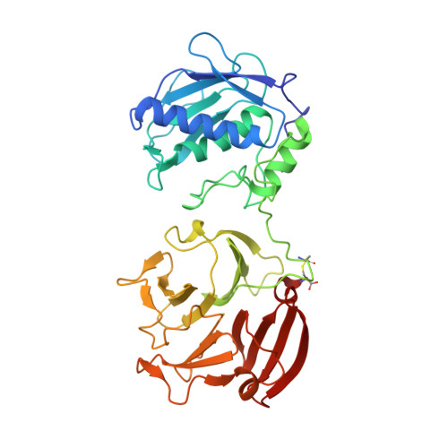
Crystal structure of full-length human collagenase 3 (MMP-13) with peptides in the active site defines exosites in the catalytic domain.

Stura, E.A.Visse, R.Cuniasse, P.Dive, V.Nagase, H.

(2013) FASEB J 27: 4395-4405

  • DOI: https://doi.org/10.1096/fj.13-233601
  • Primary Citation Related Structures: 
    4FU4, 4FVL, 4G0D

  • PubMed Abstract: 

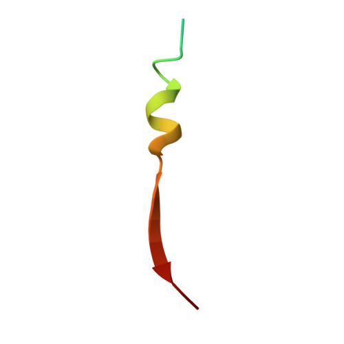
    Matrix metalloproteinase (MMP)-13 is one of the mammalian collagenases that play key roles in tissue remodelling and repair and in progression of diseases such as cancer, arthritis, atherosclerosis, and aneurysm. For collagenase to cleave triple helical collagens, the triple helical structure has to be locally unwound before hydrolysis, but this process is not well understood. We report crystal structures of catalytically inactive full-length human MMP-13(E223A) in complex with peptides of 14-26 aa derived from the cleaved prodomain during activation. Peptides are bound to the active site of the enzyme by forming an extended β-strand with Glu(40) or Tyr(46) inserted into the S1' specificity pocket. The structure of the N-terminal part of the peptides is variable and interacts with different parts of the catalytic domain. Those areas are designated substrate-dependent exosites, in that they accommodate different peptide structures, whereas the precise positioning of the substrate backbone is maintained in the active site. These modes of peptide-MMP-13 interactions have led us to propose how triple helical collagen strands fit into the active site cleft of the collagenase.


  • Organizational Affiliation
    • 2H.N., Kennedy Institute of Rheumatology, Nuffield Department of Orthopaedics, Rheumatology, and Musculoskeletal Sciences, University of Oxford, Roosevelt Drive, Oxford, OX3 7FY, UK. hideaki.nagase@kennedy.ox.ac.uk.

Macromolecule Content 

  • Total Structure Weight: 187.08 kDa 
  • Atom Count: 14,137 
  • Modeled Residue Count: 1,563 
  • Deposited Residue Count: 1,576 
  • Unique protein chains: 2

Macromolecules

Find similar proteins by:|  3D Structure
Entity ID: 1
MoleculeChains  Sequence LengthOrganismDetailsImage
Collagenase 3
A, B, C, D
368Homo sapiensMutation(s): 1 
Gene Names: MMP13
EC: 3.4.24
UniProt & NIH Common Fund Data Resources
Find proteins for P45452 (Homo sapiens)
Explore P45452 
Go to UniProtKB:  P45452
PHAROS:  P45452
GTEx:  ENSG00000137745 
Entity Groups
Sequence Clusters30% Identity50% Identity70% Identity90% Identity95% Identity100% Identity
UniProt GroupP45452
Sequence Annotations
Expand
Reference Sequence
Find similar proteins by:|  3D Structure
Entity ID: 2
MoleculeChains  Sequence LengthOrganismDetailsImage
Collagenase 3, pro-domain peptideE [auth W],
F [auth X],
G [auth Y],
H [auth Z]
26Homo sapiensMutation(s): 0 
Gene Names: MMP13
EC: 3.4.24
UniProt & NIH Common Fund Data Resources
Find proteins for P45452 (Homo sapiens)
Explore P45452 
Go to UniProtKB:  P45452
PHAROS:  P45452
GTEx:  ENSG00000137745 
Entity Groups
Sequence Clusters30% Identity50% Identity70% Identity90% Identity95% Identity100% Identity
UniProt GroupP45452
Sequence Annotations
Expand
Reference Sequence

Small Molecules

Ligands 6 Unique
IDChains Name / Formula / InChI Key2D Diagram3D Interactions
PEG

Query on PEG


Download:Ideal Coordinates CCD File
BA [auth A],
MA [auth B],
OA [auth B],
PA [auth B]
DI(HYDROXYETHYL)ETHER
C4 H10 O3
MTHSVFCYNBDYFN-UHFFFAOYSA-N
GOL

Query on GOL


Download:Ideal Coordinates CCD File
HB [auth C],
IB [auth C],
NA [auth B],
RA [auth B],
RB [auth C]
GLYCEROL
C3 H8 O3
PEDCQBHIVMGVHV-UHFFFAOYSA-N
PGO

Query on PGO


Download:Ideal Coordinates CCD File
AA [auth A]
DC [auth D]
EC [auth D]
FC [auth D]
GB [auth C]
AA [auth A],
DC [auth D],
EC [auth D],
FC [auth D],
GB [auth C],
GC [auth D],
HC [auth D],
IC [auth D],
JB [auth C],
JC [auth D],
KB [auth C],
KC [auth D],
LB [auth C],
LC [auth D],
MB [auth C],
MC [auth D],
NB [auth C],
NC [auth D],
OB [auth C],
PB [auth C],
Q [auth A],
QA [auth B],
QB [auth C],
R [auth A],
S [auth A],
SA [auth B],
SB [auth C],
T [auth A],
TA [auth B],
TB [auth C],
U [auth A],
UA [auth B],
V [auth A],
VA [auth B],
W [auth A],
X [auth A],
Y [auth A],
Z [auth A]
S-1,2-PROPANEDIOL
C3 H8 O2
DNIAPMSPPWPWGF-VKHMYHEASA-N
ZN

Query on ZN


Download:Ideal Coordinates CCD File
CA [auth B]
DA [auth B]
I [auth A]
J [auth A]
UB [auth D]
CA [auth B],
DA [auth B],
I [auth A],
J [auth A],
UB [auth D],
VB [auth D],
WA [auth C],
XA [auth C]
ZINC ION
Zn
PTFCDOFLOPIGGS-UHFFFAOYSA-N
CA

Query on CA


Download:Ideal Coordinates CCD File
AB [auth C]
AC [auth D]
BB [auth C]
CB [auth C]
EA [auth B]
AB [auth C],
AC [auth D],
BB [auth C],
CB [auth C],
EA [auth B],
EB [auth C],
FA [auth B],
FB [auth C],
GA [auth B],
HA [auth B],
IA [auth B],
K [auth A],
KA [auth B],
L [auth A],
M [auth A],
N [auth A],
O [auth A],
WB [auth D],
XB [auth D],
YA [auth C],
YB [auth D],
ZA [auth C],
ZB [auth D]
CALCIUM ION
Ca
BHPQYMZQTOCNFJ-UHFFFAOYSA-N
CL

Query on CL


Download:Ideal Coordinates CCD File
BC [auth D]
CC [auth D]
DB [auth C]
JA [auth B]
LA [auth B]
BC [auth D],
CC [auth D],
DB [auth C],
JA [auth B],
LA [auth B],
OC [auth Y],
P [auth A],
PC [auth Z]
CHLORIDE ION
Cl
VEXZGXHMUGYJMC-UHFFFAOYSA-M

Experimental Data & Validation

Experimental Data

  • Method: X-RAY DIFFRACTION
  • Resolution: 2.54 Å
  • R-Value Free:  0.241 (Depositor), 0.243 (DCC) 
  • R-Value Work:  0.169 (Depositor), 0.175 (DCC) 
  • R-Value Observed: 0.172 (Depositor) 
Space Group: P 1 21 1
Unit Cell:
Length ( Å )Angle ( ˚ )
a = 101.26α = 90
b = 105.9β = 102.11
c = 101.18γ = 90
Software Package:
Software NamePurpose
DNAdata collection
MOLREPphasing
PHENIXrefinement
XDSdata reduction
XDSdata scaling

Structure Validation

View Full Validation Report


Entry History 

Deposition Data

Revision History  (Full details and data files)

  • Version 1.0: 2013-08-21
    Type: Initial release
  • Version 1.1: 2014-02-05
    Changes: Database references
  • Version 1.2: 2023-09-13
    Changes: Data collection, Database references, Derived calculations, Refinement description
  • Version 1.3: 2024-11-20
    Changes: Structure summary