Structure-based design of pyridopyrimidinediones as dipeptidyl peptidase IV inhibitors.
Lam, B., Zhang, Z., Stafford, J.A., Skene, R.J., Shi, L., Gwaltney, S.L.(2012) Bioorg Med Chem Lett 22: 6628-6631
- PubMed: 23025999 Search on PubMed
- DOI: https://doi.org/10.1016/j.bmcl.2012.08.110
- Primary Citation Related Structures:
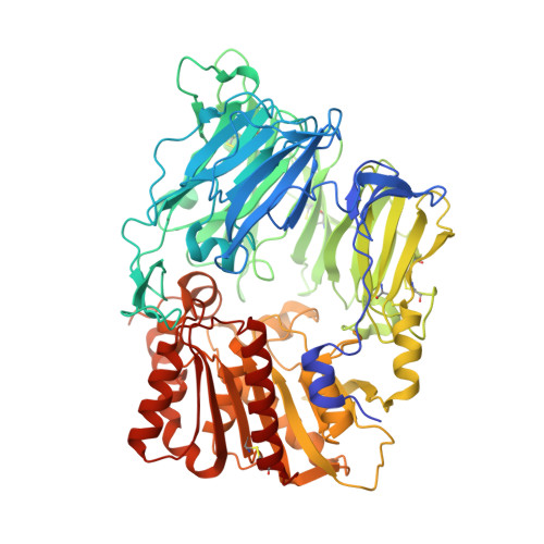
4G1F - PubMed Abstract:
Dipeptidyl peptidase IV (DPP-4) inhibitors have been shown to enhance GLP-1 levels and thereby improve hyperglycemia in type II diabetes. From a small fragment hit, using structure-based design, we have discovered a new class of non-covalent, potent and selective DPP-4 inhibitors.
- Takeda California, 10410 Science Center Drive, San Diego, CA 92121, USA. blam@takedasd.com
Organizational Affiliation: