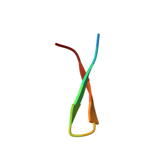
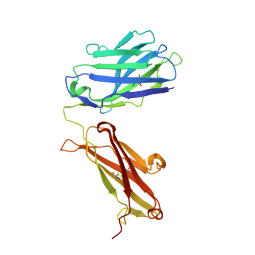
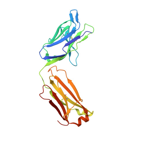
4G6A

Structure of the Hepatitis C virus envelope glycoprotein E2 antigenic region 412-423 bound to the broadly neutralizing antibody AP33


Experimental Data Snapshot

  • Method: X-RAY DIFFRACTION
  • Resolution: 2.50 Å
  • R-Value Free: 0.220 
  • R-Value Work: 0.178 
  • R-Value Observed: 0.180 

Starting Model: experimental
View more details

wwPDB Validation   3D Report Full Report


This is version 1.3 of the entry. See complete history


Literature

Structure of Hepatitis C Virus Envelope Glycoprotein E2 Antigenic Site 412 to 423 in Complex with Antibody AP33.

Kong, L.Giang, E.Nieusma, T.Robbins, J.B.Deller, M.C.Stanfield, R.L.Wilson, I.A.Law, M.

(2012) J Virol 86: 13085-13088

  • DOI: https://doi.org/10.1128/JVI.01939-12
  • Primary Citation of Related Structures:  
    4G6A

  • PubMed Abstract: 

    We have determined the crystal structure of the broadly neutralizing antibody (bnAb) AP33, bound to a peptide corresponding to hepatitis C virus (HCV) E2 envelope glycoprotein antigenic site 412 to 423. Comparison with bnAb HCV1 bound to the same epitope reveals a different angle of approach to the antigen by bnAb AP33 and slight variation in its β-hairpin conformation of the epitope. These structures establish two different modes of binding to E2 that antibodies adopt to neutralize diverse HCV.


  • Organizational Affiliation

    Department of Molecular Biology, The Scripps Research Institute, La Jolla, California, USA.


Macromolecules

Find similar proteins by:  Sequence   |   3D Structure  

Entity ID: 1
MoleculeChains Sequence LengthOrganismDetailsImage
E2 peptideA [auth B],
B [auth A]
13Hepacivirus hominisMutation(s): 0 
UniProt
Find proteins for Q9YK84 (Hepacivirus hominis)
Explore Q9YK84 
Go to UniProtKB:  Q9YK84
Entity Groups  
Sequence Clusters30% Identity50% Identity70% Identity90% Identity95% Identity100% Identity
UniProt GroupQ9YK84
Sequence Annotations
Expand
  • Reference Sequence
Find similar proteins by:  (by identity cutoff)  |  3D Structure
Entity ID: 2
MoleculeChains Sequence LengthOrganismDetailsImage
AP33 Heavy ChainC,
E [auth H]
224Homo sapiensMutation(s): 0 
Entity Groups  
Sequence Clusters30% Identity50% Identity70% Identity90% Identity95% Identity100% Identity
Sequence Annotations
Expand
  • Reference Sequence
Find similar proteins by:  (by identity cutoff)  |  3D Structure
Entity ID: 3
MoleculeChains Sequence LengthOrganismDetailsImage
AP33 Light chainD,
F [auth L]
218Homo sapiensMutation(s): 0 
Entity Groups  
Sequence Clusters30% Identity50% Identity70% Identity90% Identity95% Identity100% Identity
Sequence Annotations
Expand
  • Reference Sequence
Experimental Data & Validation

Experimental Data

  • Method: X-RAY DIFFRACTION
  • Resolution: 2.50 Å
  • R-Value Free: 0.220 
  • R-Value Work: 0.178 
  • R-Value Observed: 0.180 
  • Space Group: P 1 21 1
Unit Cell:
Length ( Å )Angle ( ˚ )
a = 44.875α = 90
b = 191.804β = 111.08
c = 59.374γ = 90
Software Package:
Software NamePurpose
Blu-Icedata collection
PHASERphasing
PHENIXrefinement
HKL-2000data reduction
SCALEPACKdata scaling

Structure Validation

View Full Validation Report


Entry History 

Deposition Data

Revision History  (Full details and data files)

  • Version 1.0: 2012-09-26
    Type: Initial release
  • Version 1.1: 2012-11-14
    Changes: Database references
  • Version 1.2: 2023-09-13
    Changes: Data collection, Database references, Refinement description
  • Version 1.3: 2024-10-30
    Changes: Structure summary