4GLY | pdb_00004gly

Human urokinase-type plasminogen activator uPA in complex with the two-disulfide bridge peptide UK504


Experimental Data Snapshot

  • Method: X-RAY DIFFRACTION
  • Resolution: 1.52 Å
  • R-Value Free: 
    0.204 (Depositor), 0.217 (DCC) 
  • R-Value Work: 
    0.128 (Depositor) 
  • R-Value Observed: 
    0.132 (Depositor) 

Starting Model: experimental
View more details

wwPDB Validation 3D Report Full Report

Validation slider image for 4GLY

This is version 1.3 of the entry. See complete history

Literature

Bicyclic Peptide Ligands Pulled out of Cysteine-Rich Peptide Libraries.

Chen, S.Rentero Rebollo, I.Buth, S.A.Morales-Sanfrutos, J.Touati, J.Leiman, P.G.Heinis, C.

(2013) J Am Chem Soc 135: 6562-6569

  • DOI: https://doi.org/10.1021/ja400461h
  • Primary Citation Related Structures: 
    4GLY

  • PubMed Abstract: 

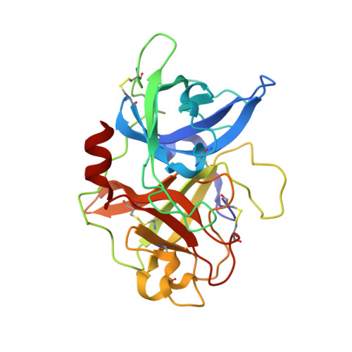
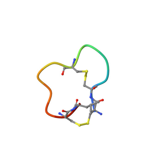
    Bicyclic peptide ligands were found to have good binding affinity and target specificity. However, the method applied to generate bicyclic ligands based on phage-peptide alkylation is technically complex and limits its application to specialized laboratories. Herein, we report a method that involves a simpler and more robust procedure that additionally allows screening of structurally more diverse bicyclic peptide libraries. In brief, phage-encoded combinatorial peptide libraries of the format X(m)CX(n)CX(o)CX(p) are oxidized to connect two pairs of cysteines (C). This allows the generation of 3 × (m + n + o + p) different peptide topologies because the fourth cysteine can appear in any of the (m + n + o + p) randomized amino acid positions (X). Panning of such libraries enriched strongly peptides with four cysteines and yielded tight binders to protein targets. X-ray structure analysis revealed an important structural role of the disulfide bridges. In summary, the presented approach offers facile access to bicyclic peptide ligands with good binding affinities.


  • Organizational Affiliation
    • Institute of Chemical Sciences and Engineering, Ecole Polytechnique Fédérale de Lausanne, CH-1015 Lausanne, Switzerland.

Macromolecule Content 

  • Total Structure Weight: 29.96 kDa 
  • Atom Count: 2,413 
  • Modeled Residue Count: 259 
  • Deposited Residue Count: 259 
  • Unique protein chains: 2

Macromolecules

Find similar proteins by:|  3D Structure
Entity ID: 1
MoleculeChains  Sequence LengthOrganismDetailsImage
Urokinase-type plasminogen activator245Homo sapiensMutation(s): 2 
Gene Names: PLAU
EC: 3.4.21.73
UniProt & NIH Common Fund Data Resources
Find proteins for P00749 (Homo sapiens)
Explore P00749 
Go to UniProtKB:  P00749
PHAROS:  P00749
GTEx:  ENSG00000122861 
Entity Groups
Sequence Clusters30% Identity50% Identity70% Identity90% Identity95% Identity100% Identity
UniProt GroupP00749
Sequence Annotations
Expand
Reference Sequence
Find similar proteins by:  Sequence   |   3D Structure  
Entity ID: 2
MoleculeChains  Sequence LengthOrganismDetailsImage
BICYCLIC PEPTIDE INHIBITOR UK50414synthetic constructMutation(s): 0 

Small Molecules

Ligands 5 Unique
IDChains Name / Formula / InChI Key2D Diagram3D Interactions
P6G

Query on P6G


Download:Ideal Coordinates CCD File
L [auth A]HEXAETHYLENE GLYCOL
C12 H26 O7
IIRDTKBZINWQAW-UHFFFAOYSA-N
SO4

Query on SO4


Download:Ideal Coordinates CCD File
C [auth A],
D [auth A],
J [auth A],
K [auth A]
SULFATE ION
O4 S
QAOWNCQODCNURD-UHFFFAOYSA-L
GOL

Query on GOL


Download:Ideal Coordinates CCD File
M [auth A]GLYCEROL
C3 H8 O3
PEDCQBHIVMGVHV-UHFFFAOYSA-N
CL

Query on CL


Download:Ideal Coordinates CCD File
F [auth A],
H [auth A],
I [auth A]
CHLORIDE ION
Cl
VEXZGXHMUGYJMC-UHFFFAOYSA-M
NA

Query on NA


Download:Ideal Coordinates CCD File
E [auth A],
G [auth A]
SODIUM ION
Na
FKNQFGJONOIPTF-UHFFFAOYSA-N

Biologically Interesting Molecules (External Reference) 

1 Unique

Experimental Data & Validation

Experimental Data

  • Method: X-RAY DIFFRACTION
  • Resolution: 1.52 Å
  • R-Value Free:  0.204 (Depositor), 0.217 (DCC) 
  • R-Value Work:  0.128 (Depositor) 
  • R-Value Observed: 0.132 (Depositor) 
Space Group: H 3
Unit Cell:
Length ( Å )Angle ( ˚ )
a = 120.8α = 90
b = 120.8β = 90
c = 43.08γ = 120
Software Package:
Software NamePurpose
REFMACrefinement
MOSFLMdata reduction
SCALAdata scaling
REFMACphasing

Structure Validation

View Full Validation Report


Entry History 

Deposition Data

Revision History  (Full details and data files)

  • Version 1.0: 2013-05-15
    Type: Initial release
  • Version 1.1: 2017-11-15
    Changes: Refinement description
  • Version 1.2: 2023-09-13
    Changes: Data collection, Database references, Derived calculations, Refinement description
  • Version 1.3: 2024-11-06
    Changes: Structure summary